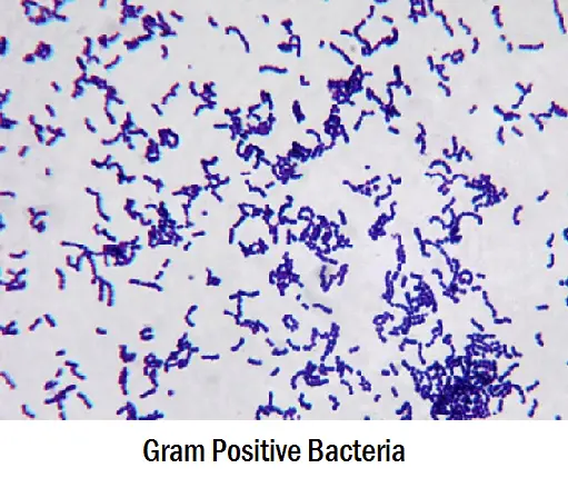
Bacteria are a huge group of single-celled microscopic organisms categorized as prokaryotic cells (lack true nucleus). Their inner structure is simple composing mainly of capsule, cellular wall, cytoplasm, ribosomes, DNA, pili, and flagellum. Bacteria can either be a gram-positive or gram-negative, and to find it out, a gram-staining technique has to be used.
Gram Staining technique is the most important and widely used microbiological differential staining technique. It categorizes bacteria according to their Gram character (Gram positive or Gram negative).
Along with their staining characteristics, Gram Positive and Gram Negative bacteria differ from each other in various aspects which are listed below :
Differences Between Gram Positive and Gram Negative Bacteria
| S.N. | Characteristics | Gram Positive | Gram Negative |
|---|---|---|---|
| 1. | Gram Reaction | Retain crystal violet dye and stain blue or purple.  | Accept safranin and stain pink or red |
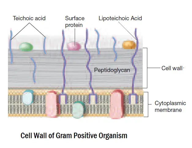
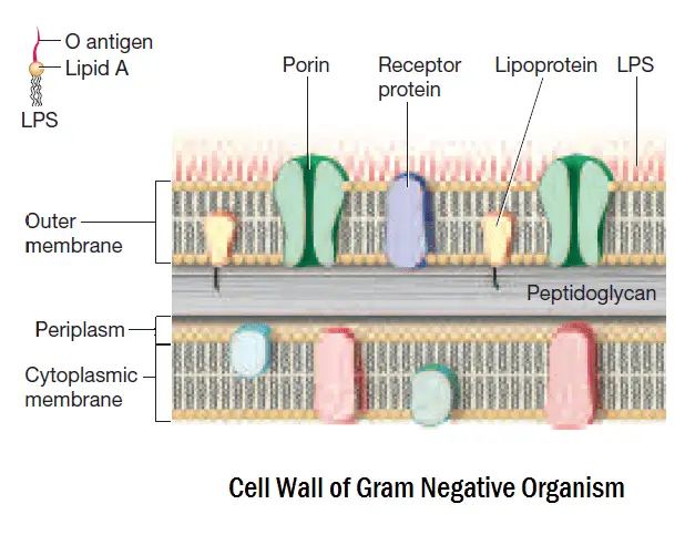
| 2. | Cell Wall Structure | Structure of Gram Positive cell wall : | Structure of Gram Negative cell wall : |
| 3. | Cell Wall Thickness | Thick (20-80 nm) | Thin (8-10 nm) |
| 4. | Chemical Composition of Cell Wall | Peptidoglycan, Teichoic acid Lipotechoic acid | Lipopolysaccharide, Lipoproteins and Peptidoglycans |
| 5. | Peptidoglycan Layer | Thick (Multilayered) | Thin (Single layered) |
| 6. | Teichoic Acid in Cell Wall | Present | Absent |
| 7. | Lipopolysaccharide Layer (Outer Layer) | Absent | Present |
| 8. | Lipid Content | Absent or lower content of lipids than Gram Negative bacteria | Contains higher content of lipids than Gram positive bacteria (due to presence of outer membrane) |
| 9. | Porin Proteins | Absent | Present |
| 10. | Flagellar Structure | 2 rings in basal body | 4 rings in basal body |
| 11. | Periplasmic Space | Absent | Present |
| 12. | Mesosome | More prominent | Less prominent |
| 13. | Ratio of RNA:DNA | 8:1 | Almost 1 |
| 14. | Toxins Produced | Primarily Exotoxins | Endotoxins and Exotoxins (Primarily Endotoxins) |
| 15. | Resistance to Physical Disruption | High | Low |
| 16. | Cell Wall Disruption by Lysozyme | High. After digestion of peptidoglycan layer, Gram +ve bacteria become Protoplast. | Low. After digestion of peptidoglycan layer, Gram -ve bacteria become Spheroplast. |
| 17. | Susceptibility to Penicilin and Sulphonamide | High | Low |
| 18. | Susceptibility to Streptomycin, Chloramphenicol and Tetracycline | Low | High |
| 19. | Inhibition by Basic Dyes | High | Low |
| 20. | Susceptibility to ionic detergents | High | Low |
| 21. | Resistance to Sodium Azide | High | Low |
| 22. | Resistance to Drying | High | Low |
| 23. | Isoelectric Range pH | 2.5-4.0 | 4.5-5.5 |
| 24. | Nutritional Requirements | Relatively Complex | Relatively Simple |
| 25. | Rendering | They can rendered Gram -ve by increasing acidity | They can rendered Gram +ve by increasing alkalinity |
| 26. | Morphology | Usually cocci or spore forming rods (exception : Lactobacillus and Corynebacterium) | Usually non-spore forming rods (Exception : Neisseria) |
| 27. | Examples | Staphylococcus Streptococcus Bacillus Clostridium Nocardia Propionibacterium Enterococcus Corynebacterium Listeria Lactobacillus Gardnerella | Escherichia Salmonella Klebsiella Proteus Helicobacter Hemophilus Vibrio Shigella Neisseria Enterobacter Pseudomonas |
Frequently Asked Questions
Q 1. What are the examples of gram-positive bacteria?
Some of the common gram-positive bacteria you will encounter in the healthcare setting as they are the leading causes of clinical infections are:
1. Staphylococcus
2. Streptococcus
3. Bacillus
4. Clostridium
5. Nocardia
6. Propionibacterium
7. Enterococcus
8. Corynebacterium
9. Listeria
10. Lactobacillus
11. Gardnerella
1. Staphylococcus
2. Streptococcus
3. Bacillus
4. Clostridium
5. Nocardia
6. Propionibacterium
7. Enterococcus
8. Corynebacterium
9. Listeria
10. Lactobacillus
11. Gardnerella
Q 2. Which is more harmful- gram-positive bacteria or gram-negative bacteria?
Of the two, gram-negative bacteria are more harmful as their outer membranes are protected by a slim layer hiding antigens present in the cell. If the infection is caused by gram-negative bacteria, it would require a strong dose of antibiotics and strict compliance to the course of treatment to thoroughly get rid of the harmful bacteria.
Q 3. Is it easier to kill gram-positive bacteria?
Of the two, gram-negative bacteria are more harmful as their outer membranes are protected by a slim layer hiding antigens present in the cell. If the infection is caused by gram-negative bacteria, it would require a strong dose of antibiotics and strict compliance to the course of treatment to thoroughly get rid of the harmful bacteria.
Q 4. What infections are caused by gram-positive bacteria?
Some of the infections caused by gram-positive bacteria include the following:
1. Urinary tract infection
2. Tuberculosis
3. Diphtheria
1. Urinary tract infection
2. Tuberculosis
3. Diphtheria
Q 5. What infections are caused by gram-negative bacteria?
Some of the infections caused by gram-negative bacteria are:
– Bloodstream infections
– Pneumonia
– Meningitis
– Bloodstream infections
– Pneumonia
– Meningitis
Q 6. Is E coli gram positive?
No. It is a rod-shaped gram-negative bacterium.
Q 7. What kills gram negative bacteria?
Gram-negative bacteria are more difficult to destroy than gram-positive. The most effective approach is to use a combination therapy, especially antibiotics with dual-mechanism action.
Q 8. Are gram negative bacteria curable?
Yes. It needs a strong dose of antibiotic and combination treatment to thoroughly destroy the pathogen.

figure of gram negative and positive bacteria is given incorrect.please have a look…
Figure gram positive and negative walls is just confusing please take a look.
The pics for gram neg and gram pos cell walls need to be swapped! Just thought I’d let you know. 😉
where can I get ur pdf filed to download ?