Gram-positive bacteria are present everywhere, but the hospitalized patients have a potential threat because of their weak immune system.
Gram-positive bacteria give positive results on the Gram stain technique, hence named gram-positive bacteria.
These bacteria retain the crystal violet dye and appear purple-colored when viewed under a microscope on a Gram stain test. Mycobacterium, Streptococci, Staphylococci, and Nocardia are some examples of gram-positive bacteria.
Characteristics of Gram-positive bacteria
Gram-positive bacteria generally have the following characteristics
- Gram-positive bacteria have thick peptidoglycan layer
- Gram-positive bacteria don’t have an outer membrane
- These have cytoplasmic lipid membrane
- Gram-positive bacteria have more teichoic acids and low lipids
- These have cilia and flagella which helps in locomotion
- These have a much smaller volume of periplasm than that in gram-negative bacteria.
Cell wall composition
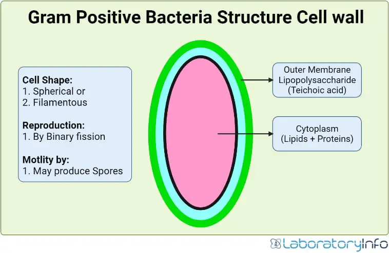
The cell wall of Gram-positive bacteria is a complex structure that consists of a thick peptidoglycan layer. This layer of peptidoglycan surrounds the cytoplasmic membrane. It includes teichoic acids, polysaccharides, and proteins.
Peptidoglycan
Peptidoglycan is the main component of Gram-positive bacterial cell walls. It is a permeable and cross-linked organic polymer composed of peptidic and glycan chains. The glycan chains are alternating N-acetylglucosamine and N-acetylmuramic acid linked through β-1,4 bonds. Peptidic chains are linked to the lactyl group through a covalent bond. Peptidoglycan provides the shape and strength to the cell wall and protects the cell from the environment.
Teichoic acid
Teichcoic acid is an anionic polymer made of the repeating units of alditol-phosphate. Teichoic acid plays an essential role in cell wall functionality. It helps control autolysins. It also helps maintain bacterial cell morphology, recognize bacteriophages, and interact with the host immune system.
Polysaccharides
Gram-positive bacterial cell walls also have polysaccharides in their cell wall. The three main groups are exopolysaccharides, capsular polysaccharides, and cell wall polysaccharides.
Classification
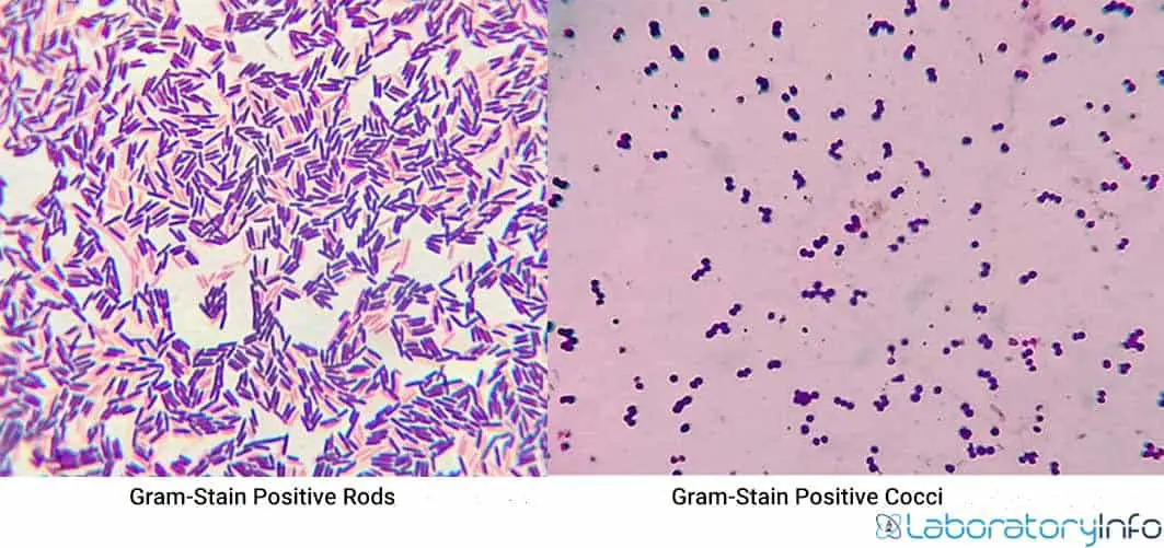
- There are two main types of Gram-positive bacteria; cocci and bacilli (rods).
- The cocci are oval or spherical in shape.
- These have further arrangements like diplococci, streptococci, staphylococci, tetrads, and sarcina. The bacilli are rod-shaped bacteria and are found as diplobacilli, streptobacilli, and coccobacillus.
Gram positive bacteria list

Differences between cocci and bacilli
The differences between cocci and bacilli are following
| Cocci | Bacilli |
| These are either spherical, oval, or kidney-shaped | These are either rod, vibrio, or spindle-shaped |
| One axis of bacterium is same as the other | One axis of bacterium is longer than the other |
| Both Gram-positive and Gram-negative | Mostly Gram-positive |
| Mostly anaerobe | Either aerobe or anaerobe |
| Examples are Neisseria, Streptococcus, Staphylococcus, etc | Examples are Clostridium, Nocardia, Escherichia, etc |
Gram-positive cocci
Bacterium that is spherical, ovoid or round-shaped is called coccus. Diplococci is a pair of cocci and streptococci is a chain of cocci. Staphylococci are Irregular clusters of cocci and four cocci arranged in the same plane are called tetrad.
Sarcina is the cuboidal arrangement of eight cocci. Gram-positive cocci are divided into; staphylococci and streptococci.
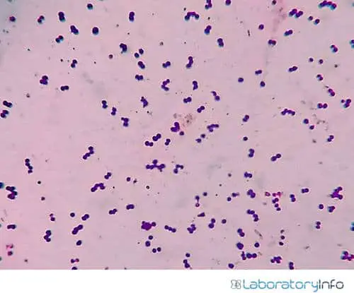
Image Source: Wikimedia CC
- Staphylococci bacteria are divided into coagulase-positive and coagulase-negative species. Staph aureus is an example of a coagulase-positive organism. Staph epidermidis and Staph saprophyticus are coagulase-negative pathogens.
- Streptococci bacteria are further divided into Strep. pyogenes (also called Group A), Strep. agalactiae (also called Group B), enterococci (also called Group D), Strep pneumonia, and Strep viridans.
Gram-positive bacilli (rods)
Gram-positive bacilli are rod-shaped and mostly arranged as a single bacterium. Vibrio is a single curved rod. If the two bacilli are arranged side by side, these are called diplobacilli. Streptobacilli are the chains of bacteria and Coccobacillus is a short rod-shaped bacteria.
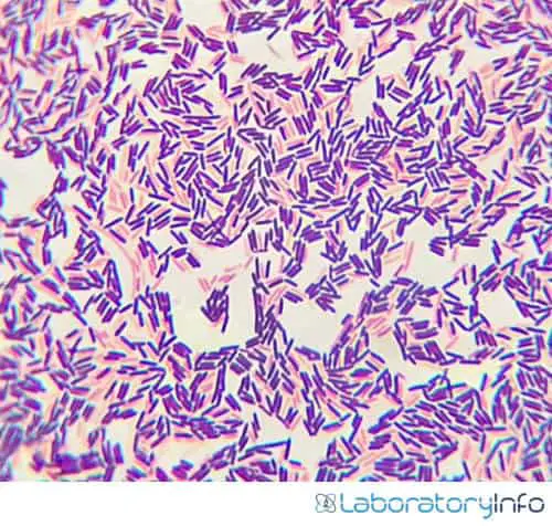
Image Source: Wikimedia CC
- It is divided according to their ability to produce spores.
- Bacillus and Clostridia are grouped as spore-forming bacilli (rods), while Listeria and Corynebacterium don’t form spores.
- The spores produced by the spore-forming rods can survive in the environment for many years.
Antibiotics for Gram-positive bacteria
Gram-positive bacteria are among the most common infectious causes. The clinical conditions may range from mild skin to sepsis. Several antibiotics exist for treating the disorders caused by these bacteria.
- Penicillin, cloxacillin, and erythromycin cover almost 90 % of Gram-positive infections.
- Quinupristin/dalfopristin and linezolid are effective against some strains of gram-positive bacteria that are resistant to vancomycin.
- Vancomycin and teicoplanin are glycopeptides. These are bactericidal antibiotics with activity against Gram-positive bacteria only. These are used for the treatment of SSTIs.
- Daptomycin is a broadspectrum cyclic lipopeptide. It is active against Gram-positive bacteria, including GRE and MRSA. Daptomycin is being used for SSTIs, right-sided infective endocarditis, and bacteremia secondary to S.aureus.
- Ceftaroline and ceftobiprole are novel fifth-generation cephalosporins with a unique bactericidal activity spectrum. Ceftaroline is being used to treat complicated SSTIs and community-acquired pneumonia (CAP) in adults.
Frequently Asked Questions
Q1. What is Gram Staining?
Gram staining is a widely used standard procedure in microbiology. It is used to classify the bacteria according to their cell wall composition.
The Danish Bacteriologist, Hans Christian Gram, developed this technique in 1884. He devised a method to identify whether or not a bacteria had a peptidoglycan wall.
This technique classifies the bacteria into two major groups; Gram-positive and Gram-negative bacteria. Gram-staining is also a valuable diagnostic tool in both clinical and research settings.
Q2. How is Gram-staining performed?
Gram-staining is a test to classify the bacteria. It identifies whether bacteria have a peptidoglycan layer or not. We have to introduce a dye to bacteria. If bacteria have a thick peptidoglycan cell wall, it absorbs the stain and turns purple-color. And we can say that it’s positive for peptidoglycan. If it’s not turned into purple color, the test is negative for peptidoglycan.
Q.3 What is the use of Gram-staining?
The technique is widely used to classify bacteria based on their cell wall structure. It classifies the bacteria into gram-positive and gram-negative bacteria.
Q.4 What are the specimens used for Gram-staining?
The specimens used for this technique follow
1. Blood
2. Tissue
3. Stool
4. Urine
5. Sputum
Q.5 Define resident flora?
The microorganisms living harmlessly on the body are called resident flora.
Q.6 What is a peptidoglycan wall?
Bacterial cells are enclosed by a peptidoglycan wall, a mesh-like layer of sugars and amino acids. This mesh-like structure is semi-permeable.
Q.7 What is the importance of the cell wall of Gram-positive bacteria?
The outer layer of bacteria is essential because
1. This mesh-like structure protects the cellular contents from external environmental factors.
2. It also maintains cell shape and integrity during growth and division.
3. Maintains the inside pressure of the bacterial cell.
4. Permeable to small molecules and maintains cellular metabolism.
5. It prevents larger and potentially toxic substances from entering the cell.
Q.8 Do Gram-positive bacteria have capsule?
The bacterial capsule is mostly made of polysaccharides. It can be found in both Gram-positive and Gram-negative bacteria.
Q.9 What is the difference between Staphylococci and Streptococci?
Staphylococci
1. These are catalase-positive.
2. Grows in clusters.
Streptococci
1. These are catalase-negative.
2. Grows in chains.
Q.10 Is E.coli a Gram-positive bacteria?
No, E.coli is a Gram-negative bacteria.
Q.11 What are the examples of Gram-positive bacilli (rods)?
Examples of Gram-positive bacilli are Corneybacterium, Listeria, Nocardia, etc.
Q.12 What are the examples of Gram-positive cocci?
The examples of Gram-positive cocci are Staphylococcus aureus, S. epidermidis, Streptococcus pyogenes, S. pneumoniae, etc.
Q.13 Which antibiotic is effective for Gram-positive bacteria?
The antibiotics effective against the Gram-positive bacteria are penicillin, cloxacillin, and erythromycin cover almost 90 % of Gram-positive infections. Others are quinupristin/dalfopristin, linezolid, vancomycin, daptomycin, etc.
References
- Sizar O, Unakal CG. Gram-Positive Bacteria. [Updated 2022 Feb 14]. In: StatPearls [Internet]. Treasure Island (FL): StatPearls Publishing; 2022 Jan-.
- A. (2021, March 22). General Data Protection Regulation(GDPR) Guidelines BYJU’S. BYJUS. https://byjus.com/biology/gram-positive-bacteria/
- Chapot-Chartier, MP., Kulakauskas, S. Cell wall structure, and function in lactic acid bacteria. Microb Cell Fact 13, S9 (2014). https://doi.org/10.1186/1475-2859-13-S1-S9
- Bruslind, L. (2019, August 1). Bacteria: Cell Walls – General Microbiology. Pressbooks. https://open.oregonstate.education/generalmicrobiology/chapter/bacteria-cell-walls/
- Antimicrobial therapies for Gram-positive infections. (2021, February 12). The Pharmaceutical Journal. https://pharmaceutical-journal.com/article/research/antimicrobial-therapies-for-gram-positive-infections
