A stereo microscope is not new. It has been around for a while now. It is also called a dissecting microscope and stereoscopic.
It provides a 3-D view of the specimen. Each eye has separate objective lenses and eyepieces. What sets it apart from a compound microscope is that it has a lower magnification and longer working distance.

Image 1: A stereo microscope provides a 3D view of the specimen.
Picture Source: microscopeworld.com
History
- Cherubin d’Orleans (1613-1697) – He was a monk who designed and built the first pseudo-stereoscopic. He created a small microscope with two separate objective lenses and eyepieces.
- Charles Wheatstone (1802-1875) – He is an English scientist and inventor who first described the principle of stereoscopic vision.
- John Leonard Riddell (1807-1865) – Further advancement in stereo microscopy was made by him. In his paper, ‘On the Binocular Microscope’4, he published his finding in the Quarterly Journal of Microscopical Science.
- Horatio S. Greenough – He is an American biologist and instrument maker who developed a stereo microscope as an alternative design to CMO microscope. Today’s stereo microscope is still based on the principles of Horatio S. Greenough. (1, 2, 3, and 4)
Uses
- A stereo microscope is primarily used to view specimens like plants and animals.
- A stereo microscope is useful when working with circuits and watches.
- It can be used for microsurgery.
- It is used to view crystals. (2,3, 4, and 5)

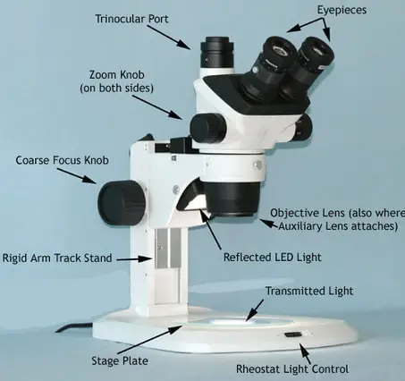
Image 2: The different parts of a stereo microscope.
Picture Source: microbehunter.com
Stereo Microscope Parts and Functions
It has three key parts namely: body, focus block, and viewing head/body. Let us take a look at the functions of every part.
- Body/viewing head – It houses the optical parts in the upper section of the microscope.
- Focus block – It attaches the head of the microscope to the stand and focuses it.
- Stand – It supports the microscope as well as houses integrated illumination. (4, 5)
Optical Parts
- Eyepiece Lenses – These are situated at the top of the microscope. a. Standard eyepiece – It has a magnifying power of 10X.
- Optional eyepieces – It has varying power ranging between 5X and 30X.
- Eyepiece tube – It keeps the eyepieces in place; situated just above the objective lens.
- Diopter adjustment ring – It enables possible inconsistencies of the eyesight in one/both eyes.
- Objective Lenses – They are the microscope
Other vital parts
- Focus – Stereo microscope has coarse focus controls.
- Working stage – it is a place where the specimen to be examined is put on.
- Stage clips – They are used only with the absence of a mechanical stage.
- Transmitted illumination – It is used to shed light on the specimen. (5, 6)
Stereo Microscope Types

Image 3: A stereo fixed microscope.
Picture Source: microscopegenius.com
Stereo fixed microscope
it is called a fixed stereo microscope because of its fixed magnification using two objective lenses. The magnification has a fixed degree and is limited by the lens capability. To increase the magnification, you need to change the eyepiece.

Image 4: A stereo turret microscope.
Picture Source: microscopeworld.com
Stereo Turret Microscope
It comes with various mountings and one of which is the turret style. This type of mounting indicates an additional objective lens which you can rotate to your viewing position.
The viewer can easily change the magnification by rotating the mounting of a turret. It is preferred by many because it is more affordable than other types of stereo microscope.

Image 5: A stereo zoom microscope.
Stereo Zoom Microscope
This is the most popular type of stereo microscope. You can easily zoom in and out to achieve the desired magnification. You can have a clear magnification by changing the eyepiece. It comes with different stands such as:
- Simple stand – It provides room for observation and repair of items.
- Boom stand – It is used for larger applications. It can be easily mounted to the floor and comes with a boom which serves as a counterweight. It is more expensive than the simple stand primarily because of the structure and functions. (5, 6, 7, and 8)
What to keep in mind when using a stereo microscope?
- Place the stereo microscope in a flat and stable surface.
- Turn on the illuminator. Put a specimen onto the stage.
- The magnification adjustment knob should be turned to the lowest power and focus the image using focus control.
- Adjust the eyepiece according to your level of comfort. Move the eyepieces closer together or far apart until such time you achieve a single field of view.
- The dioptric adjustment ring should be set to zero position.
- Always use the magnification adjustment to reach the highest magnification. The focus knob is used to focus the image being examined.
- The image could be a bit out of focus if you use the lowest magnification.
- If you want to adjust the focus, you need to use the eyepiece dioptric adjustment ring.
- If you are examining a solid or opaque object, you should use the incident light. If you are going to examine a transparent specimen, you should use the bottom light.
- A stereo microscope enables 3D view of the specimen.
- It is also known as low power/dissecting microscope.
- Some stereo microscopes are dual power, which provides two or more fixed levels of magnification.
- A stereo microscope is modular, which means that you can use the same head in conjunction with various focus blocks and stands.
- If you want to calculate the magnification of a stereo microscope, you need to multiply the eyepiece and the zoom knob. If you are using the auxiliary lens, you need to include it in the equation, which means you need to multiply the eyepiece, zoom knob, and auxiliary lens. (3, 5, 7, and 8)

Image 6: A comparison image between stereo and compound microscope.
Picture Source: stereomicroscopes.org
Differences between – Stereo vs Compound microscope
Types
- Stereo – Optical microscope
- Compound – Light microscope
Optical Path
- Stereo – It uses episcopic or reflected illumination to light up the subject/light naturally reflected from the specimen.
- Compound – It has a single optical path giving the same image in both left and right eye.
Specimens being examined
- Stereo – plants, animals, crystals, circuits, coins, jewelry, textiles, and watches.
- Compound – Bacteria, plant cells, animal cells, chromosomes, and blood counts.
Magnification
- Stereo – It presents a slightly different view to each eye.
- Compound – It magnifies in two stages.
Nature
- Stereo – It is used for checking items in which light cannot shine through. It enables you to observe the actual color of the specimen.
- Compound – It is used to check ultra-thin pieces of large objects.
Structure
- Stereo – It has two lenses aligned in a way that brings a 3D magnification on the specimen.
- Compound – It uses multiple lenses to focus the light to the viewer’s eye. Structure-wise, a compound microscope is large, heavy, and more expensive.
Amplification
- Stereo microscope – It uses low amplification.
- Compound microscope – It uses high amplification; as high as 1000X.
Working space
- Stereo microscope – Large working space
- Compound microscope – Little working space
Light
- Stereo microscope – The specimen is viewed using reflected light.
- Compound microscope – The light is transmitted through the object.
Functions
- Stereo microscope – It is used to examine the surface of solid substances.
- Compound microscope – It is used to examine minute things. (4, 6, 9, and 10)
| Points of comparison | Stereo microscope | Compound microscope |
| Types | Optical microscope | Light microscope |
| Optical path | Episcopic or reflected illumination | Single optical path |
| Specimens being examined |
|
|
| Magnification | slightly different view to each eye | magnifies in two stages |
| Nature | Checks items in which light cannot shine through. | Checks ultra-thin pieces of large objects. |
| Structure | It has two lenses aligned in a way that brings a 3D magnification on the specimen. | It uses multiple lenses to focus the light to the viewer’s eye. |
| Amplification | Low amplification | High amplification |
| Working space | Large working space | Little working space |
| Light | Reflected light | Light is transmitted through the object |
| Functions | It is used to examine the surface of solid substances. | It is used to examine minute things. (3, 6, 9, and 10) |
Stereo microscope Limiting factors
The working distance between the lens and the specimen/sample is one of the limiting factors when using a stereo microscope. If you are going to increase the magnification, the distance and the field of view will be decreased.
If the magnification is decreased, you will need to increase your working distance as well as the field of view. The more you magnify the specimen, the closer your focal point moves. Greater magnification leads to a bigger view but the aperture does not change.
As the image is enlarged, the lesser area you see under the microscope. If you are planning to buy a stereo microscope, make sure you check the magnification setting/magnification power so as to make sure that the one you are getting provides a large field of view.
You should also check the working distance and make sure it is large enough to fit your sample between the lens and the base. (3, 5, 7, and 8)
References
- https://en.wikipedia.org/wiki/Stereo_microscope
- https://www.microscope-detective.com/stereo-microscope.html
- https://www.microscopeworld.com/c-429-stereo-microscopes.aspx
- https://www.microscope.com/education-center/five-things-you-should-know/about-stereo-microscopes/
- https://www.microscope.com/stereo-microscopes/
- http://microscopy.berkeley.edu/courses/tlm/stereo/index.html
- https://www2.mrc-lmb.cam.ac.uk/microscopes4schools/microscopes1.php
- https://www.microscopemaster.com/stereo-microscope.html
- http://accu-scope.com/support/frequently-asked-questions/
- http://www.microbehunter.com/what-are-the-differences-between-stereo-and-compound-microscopes/
