A compound microscope is a laboratory instrument used to magnify the image of a small object; usually objects that cannot be seen by the naked eye.
It comes with two or more lenses, which causes it to achieve a higher level of magnification when compared with other low power microscopes. A compound microscope has the following:
- It comes with two or more convex lenses.
- One objective is used at a time.
- It produces 2-dimensional images.
- Its typical magnification is between 40x and 1000x.
- It is available in different configurations: monocular, binocular, and trinocular. (1, 2, 3, and 4)
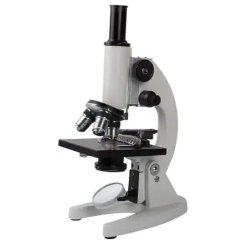
Image 1: The image is a typical compound microscope commonly found in the workplace.
Picture Source: imimg.com
Who invented the compound microscope?
The invention of the compound microscope is credited by historians to Zacharias Janssen, a Dutch spectacle maker, around 1590.
Principles of compound microscope
When a minute object is placed beyond the focus of the objective lens, a highly magnified object is formed at a distance of distinct vision from the eye close to the eye piece. A compound microscope has two convex lenses; an objective lens and eye piece.
The objective lens is placed towards the object and the eyepiece is the lens towards our eye. Both eyepiece and objective lenses have a short focal length and fitted at the free ends of two sliding tubes. (4, 5, and 6)
Compound microscope parts and magnification
A compound microscope consists of different parts and each part plays an important function. These include the following:
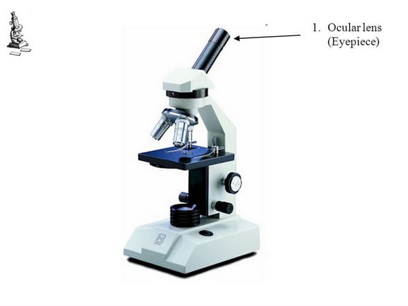
Image 2: The eyepiece/ocular lens of a compound microscope.
Picture Source: slideplayer.com
- Eyepiece/ocular lens – It is the part of the microscope that is looked through at the top. It comes with a magnification ranging between 5x and 30x.

Image 3: The head connects the eyepiece to the objective lens.
Picture Source: microscope.com
- Head (monocular/binocular) – It is the structural support of the microscope. It holds and connects the eyepiece to the objective lens.

Image 4: The objectives of a compound microscope.
Picture Source: slideplayer.com
- Objective lens – A compound microscope has three to five optical lens objectives and each comes with various magnification level (4x, 10x, 40x, and 100x). To calculate the total magnification of the microscope, all you need to do is to multiply the objective lens magnification by eyepiece magnification level.

Image 5: The arm of the compound microscope.
Picture Source: zfic.org
- Arm – it supports the head of the microscope and attach it to the base.

Image 6: The revolving nosepiece.
Picture Source: img-aws.ehowcdn.com
- Nosepiece – It holds the objective lens and attaches them to the head of the microscope. You can rotate the nosepiece to change the objective lens.

Image 7: The base is the bottom part of the microscope.
Picture Source: microscopeinternational.com
- Base – It supports the microscope and houses the illumination of the microscope.

Image 8: A sliding glass is needed and is attached to the stage using a stage clip.
Picture Source: images-na.ssl-images-amazon.com
- Sliding glass – It holds the specimen for easy viewing. It is made of thin rectangular glass.

Image 9: This is how a stage clip looks like.
Picture Source: microscope.com
- Stage clip – It clips/holds the sliding glass in place.

Image 10: The stage is where the sliding glass with a specimen is placed,
Picture Source: microscope.com
- Stage/platform – It is where the specimen or slide is placed.

Image 11: The aperture diaphragm control.
Picture Source: stevegallik.org
- Aperture – It is disc characterized by its circular opening where the illumination from the base reaches the platform stage.

Image 12: The condenser is underneath the stage.
Picture Source: microscope.com
- Abbe condenser – It is a lens that condenses the light from the base illumination and directed it to the stage.

Image 13: The coarse and fine adjustment knobs.
Picture Source: boruhealthmachine.org
- Adjustment controls/knob (coarse/fine) – It allows you to easily adjust the focus of the microscope. By adjusting the knob, you can easily increase or decrease the level of details seen when examining at the slide through the eyepiece.
- Coarse focus – Use this knob with the lowest power objective to get the subject in focus.
- Fine focus – It is the smaller of the two focus knobs. It is the commonly used focus in viewing the slides.

Image 14: The stage height adjustment is at the bottom left.
Picture Source: slideplayer.com
- Stage height adjustment – It allows you to easily adjust the placement of mechanical stage in both horizontal and vertical path. Adjusting the knob is important as it prevents the possibility of contact between the objective lens and slide containing the specimen.

Image 15: The illumination is at the center of the base.
Picture Source: microscopeinternational.com
- Illumination – It is the light used to illuminate the slide that contains the specimen. The light comes from the base of the microscope.

Image 16: Mirror sets on top of the base.
Picture Source: enasco.com
- Mirror – it reflects light into the microscope’s base.

Image 17: The field diaphragm.
Picture Source: olympus-lifescience.com
- Bottom lens/field diaphragm – it is a knob used to adjust the amount of light that gets in contact with the specimen. (5, 6, 7, and 8)
How a compound microscope works/functions?
Light begins at the base of the microscope coming from the source of illumination. It travels upward through the condenser and aperture and passes through the stage. As the light passes through, the image of the specimen on the slide is picked up by the magnification of the objective lens above it. The magnification varies. After which, the light moves to the head of the microscope reaching the eyepiece and magnified by the ocular lenses.
Basically, all the parts of the microscope work together to magnify the specimen and have a clearer view. As someone who is using the microscope, it is important to learn how to properly use and adjust the microscope.
Aside from the proper use of a microscope, it is also important to keep the microscope in perfect shape and one way of doing so is by keeping it clean. (2, 5, 8, and 9)
Why is compound microscope image inverted?
A compound microscope captures an inverted image of the specimen because every time the light passes through the lens, the image’s direction is flipped. The image always ends up inverted from the original. So, if you move the sample to the left, it moves in the right direction.

Image 18: A comparison image between a simple and compound microscope.
Picture Source: microscopeheroes.com
What is the difference between a compound microscope and a simple microscope?
Meaning
- Simple microscope – It is a convex lens of small focal length and its primary use is to see a magnified image of small objects.
- Compound microscope – It is an optical instrument consists of two convex lenses of short focal lengths primarily used for observing a highly magnified image of minute objects.
Lenses
- Simple microscope – It has a convex lens. It uses only one lens to magnify objects. An example of a simple microscope is a magnifying glass.
- Compound microscope – It has two convex lenses. It is called a compound microscope because it compounds the light as it passes through the lenses to magnify. The image of the object being viewed is enlarged because of the lens near the object. An eyepiece, an additional lens, is where real magnification takes place. The lens of the eye piece magnified the already enlarged image making it larger and clearer. (2, 4, and 6)
Focal Length
The focal length is the distance between the lens and its focus.
- Simple microscope – A simple microscope has a short focal length.
- Compound microscope – They eyepiece makes the focal length longer and more precise. The objective lens and the eyepiece make the object larger and more defined.
Magnification
- Simple microscope – It has a maximum magnifying power of 10. As with the nature of magnification, a simple microscope has a fixed magnification. It magnifies the image to a certain degree that the lens allows.
- Compound microscope – It has the maximum magnifying power of 1000. A compound microscope’s magnification can be multiplied because it has an additional lens. You can magnify to the lens the highest capacity making the image clearer and more defined. (7, 9, and 10)
Presence of condenser lens
- Simple microscope – Absent
- Compound microscope – Present
Source of light
- Simple microscope – Natural
- Compound microscope – Illuminator
Type of mirror
- Simple microscope – Concave reflecting
- Compound microscope – It has both plain and concave type mirror.
Magnification adjustment
- Simple microscope – No
- Compound microscope – Yes
Usage/application
- Simple microscope – For simple/basic use.
- Compound microscope – A compound microscope is commonly used for professional research purpose.
Check the table below for a detailed comparison between a simple microscope and a compound microscope.
| Point of comparison | Simple microscope | Compound microscope |
| Definition | A convex lens of small focal length and its primary use is to see the magnified image of small objects. | An optical instrument consists of two convex lenses of short focal lengths primarily used for observing a highly magnified image of minute objects. |
| Lenses | A convex lens with only one lens to magnify the object. | Two convex lenses |
| Focal length | short focal length | Longer and more precise |
| Magnification | Fixed magnification | Varies |
| Presence of condenser lens | Absent | Present |
| Source of light | Natural | Illuminator |
| Type of mirror | Concave reflecting | Both plain and concave |
| Magnification adjustment | No | Yes |
| Usage/application | Simple/basic use | Professional research purpose |
Compound microscope types
Compound microscopes are categorized into four types. They are the following:

Image 19: A toy compound microscope.
Picture Source: ebayimg.com
- Toy microscopes – These are compound microscopes sold in toy shops. You can easily spot a toy microscope because it comes with many accessories which include but not limited to the following:
- Slides
- Plastic pipettes
- Prepared specimens
- Tweezers
- Ridiculously high magnification
A toy microscope does not have objectives manufactured as per the 160 mm standard. It has a low resolution and it is extremely difficult to achieve focus because its parts are made of plastics.
Another distinct characteristic of the toy compound microscope is its low field of view and low brightness.

Image 20: A compound microscope typically used in schools.
Picture Source: medpro-microscope.com
Educational/student microscope
- This type of compound microscope is small, which makes it a portable device. So, students can bring it with them anytime and anywhere. Its eyepiece has 10x magnification and comes with three objectives: 4x, 10x, and 40x.
- Its light source comes from halogen or LED. Some even have a battery, which enables you to use the microscope even with no power supply. This is the best microscope for an amateur user.
- It is easy to use and adheres with the DIN standardized objectives. Its body is usually made from metal. Some educational/student compound microscopes have a condenser with a diaphragm for you to easily control the resolution, contrast, and depth of field.

Image 21: A compound microscope typically used in the laboratory setting.
Picture Source: ssl-images-amazon.com
Routine/laboratory microscope
- It is bigger in size when compared with the student microscope. It is also heavier. It is primarily used for laboratory setting. It comes with a wide body and base.
- Its distinct parts include a condenser, illumination, focus lock, mechanical stage, and a revolving nosepiece which can hold up to five objectives. It usually has a binocular head, which makes long-term observation easy.

Image 22: An example of a research compound microscope.
Picture Source: ssl-images-amazon.com
Research microscope
- This type of compound microscope is used for extensive research. It is large, heavy, quite modular, not portable, and too expensive when compared with other types of compound microscope.
- It comes with different objectives and filters. (2, 4, 7, 9, 10, and 11)
References
- https://www.microscopeinternational.com/what-is-a-compound-microscope/
- https://www.microscope.com/compound-microscope-parts/
- https://www2.mrc-lmb.cam.ac.uk/microscopes4schools/microscopes1.php
- http://microscopy.berkeley.edu/courses/tlm/cmpd/cmpd.html
- http://www.funscience.in/study-zone/Physics/OpticalInstruments/CompoundMicroscope.php#sthash.lZVVb7bU.dpbs
- http://www.math.ubc.ca/~cass/courses/m309-03a/m309-projects/yeh/micro.htm
- https://www.martinmicroscope.com/microscopes/
- https://optimaxonline.com/newsdetails.php?newsId=21
- https://courses.lumenlearning.com/ap1x94x1/chapter/the-parts-of-a-compound-microscope-and-how-to-handle-them-correctly/
- http://hyperphysics.phy-astr.gsu.edu/hbase/geoopt/micros.html
- https://staff.concord.org/~btinker/GL/web/water/using_compound_microscope.html