A microscope is one of the invaluable tools in the laboratory setting. It is used to observe things that cannot be seen by the naked eye.
From a simple microscope, there are now most advanced versions which perform a more complex function. Microscopes are specially created to magnify the image of the subject being studied.
This exercise is created to be used in homes and schools. the microscope layout, including the blank and answered versions are available as pdf downloads.
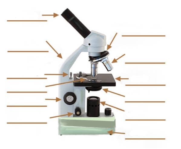
Click to Download : Label the Parts of the Microscope (A4) PDF print version.

Click to Download : Label the Parts of the Microscope with answers (A4) PDF print version.
For a thorough review of each microscope part continue reading….
A basic microscope has a single convex lens such as those found in a magnifying glass, which you can use to visualize the finest prints.
Using the magnifying glass, you can make the object appear bigger by 10 to 20 times. In the most advanced microscopes, multiple lenses are combined to get a greater magnification. A standard microscope has two lenses namely the objective lens and the eyepiece.
A light is needed to shine on the object and then reflected by the mirror into the lenses, hence, causing greater magnification. (1, 2, 3, and 4)
Let us take a look at the different parts of microscopes and their respective functions.
1. Eyepiece
it is the topmost part of the microscope. Through the eyepiece, you can visualize the object being studied. Its magnification capacity ranges between 10 and 15 times.

2. Body tube/Head
It is the structure that connects the eyepiece to the lenses.
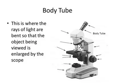
Image 2: The body tube part of a microscope is where the ray of light is bent to allow the object being viewed to enlarge by the scope.
Picture Source: slideplayer.com
3. Turret/Nose piece
It is the revolving part of the microscope. It allows the use of different types of objective lenses by simply rotating the top part of the turret. If you wish to examine the subject being studied at a different magnification, all you need to do is to rotate the top part of the turret.
Image 3: A turret is a structure found above the objective lenses which can be easily rotated so that you will be able to choose the most suited lens.

Picture Source: thorlabs.com
4. Objective lenses
A microscope usually has 3 to 4 objective lenses with varying magnifications, usually 10 to 100x.

Image 4: A closer look at the different objective lenses.
Picture Source: microscopemaster.com
5. Knobs (fine and coarse)
By adjusting the knob, you can adjust the focus of the microscope. The majority of the microscope models today have the knobs mounted on the same part of the device.
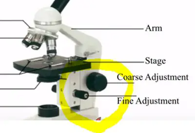
Image 5: The circled parts of the microscope are the fine and coarse adjustment knobs.
Picture Source: bp.blogspot.com
6. Stage and stage clips
You put the specimen being studied on the stage and to secure the specimen in place you will need to use the stage clips.

Image 6: A closer look at the stage and the surrounding structures as the clips and stage opening.
Picture Source: olympus-lifescience.com
7. Aperture
Examine the microscope and you will see a hole from which the light shines. The structure is called an aperture.

Image 7: A numerical aperture of a simple microscope.
Picture Source: magnet.fsu.edu
8. Illuminator
It illuminates the light to the specimen for better visualization. It is the one responsible for creating light rays for magnification.

Image 8: The encircled part of the microscope is the illuminator.
Picture Source: alicdn.com
9. Condenser
The light from the illuminator is collected by the condenser and direct it on the specimen.

Image 9: These are some of the condenser found in a microscope.
Picture Source: olympus-lifescience.com
10. Condenser focus knob
This structure helps the condenser to move up and down to better control the image of the specimen.
11. Iris diaphragm
The primary function of iris diaphragm is to control the intensity of light as well as helps focus the light on the specimen. (7, 8, 9, and 10)
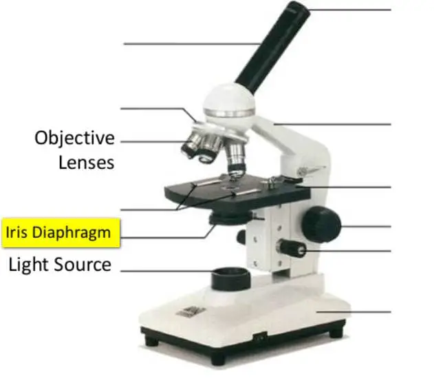
Image 11: An example of a microscope with an iris diaphragm.
Picture Source: stcgrupo.co
12. Diopter adjustment
It is used to adjust the focus on one eyepiece, which is helpful in correcting difference in vision between the two eyes.

Image 12: An eye piece with an attached diopter adjustment.
Picture Source: digital-usb-microscopes.com
13. Arm
It is a structure that connects the body tube to the microscope’s base.
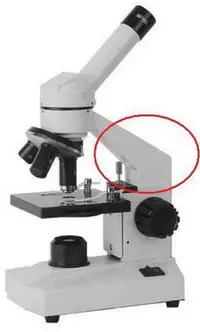
Image 13: The image above is the arm of the microscope.
Picture Source: easynotecards.com
14. Specimen/slide
It is the object being studied and is mounted on the slides, which is a sheet of glass that is flat and rectangle in shape. The specimen is put on the glass and the slide is inserted into the stage.

Image 14: The image above is an example of a glass slide where the specimen is placed.
Picture Source: indepthinfo.com
15. Stage control/stage height adjustment
It is the part of the microscope that allows the stage to move left and right and up and down.

Image 15: The stage control feature of the microscope can be adjusted to move the stage to the desired direction.
Picture Source: olympus-lifescience.com
16. On and off switch
It is located on the base of the microscope. Turn the switch to turn the illuminator on and off.

Image 16: A closer look at the on and off switch of a microscope.
Picture Source: cas.miamioh.edu
17. Base
it is found in the bottom part of the microscope where the illuminator is located. The base supports the entire structure of the microscope.

Image 17: The base of the microscope provides stability for the entire structures.
Picture Source: miamioh.edu
References
- https://www.microscopeworld.com/t-parts.aspx
- https://microscope-microscope.org/microscope-info/microscope-parts/
- https://www.microscopemaster.com/parts-of-a-compound-microscope.html
- https://www.amscope.com/microscope-parts-and-functions/
- https://www.microscope.com/compound-microscope-parts
- https://sciencing.com/parts-microscope-uses-7431114.html
- https://microbenotes.com/parts-of-a-microscope/
- http://enfo.agt.bme.hu/drupal/en/node/8862
- https://www.grainger.com/know-how/equipment-information/kh-microscope-types-parts-terms
- https://www.pobschools.org/cms/lib/NY01001456/Centricity/Domain/349/TheMicroscope-howtouse.pdf