In the cells, you will find tiny little projections called microvilli. They can be on their own or with villi. In the small intestines, you will find tiny folds that look like fingers or referred to as villi.
In every villus, there are smaller folds called microvilli, which have their own plasma membrane covering them. Inside the microvilli is a cytoplasm or cellular fluid and microfilaments giving microvilli the structure it needs.

Image 1: A closer look at the structure of microvilli.
Picture Source: britannica.com
Where are microvilli located?
They are abundant in the small intestines, egg cells’ surface, and in the white blood cells. In the intestines, they exist together with the villi and their function is to expand the surface area of the intestines so that they will be able to absorb more nutrients and materials.
In the egg cells, microvilli help anchor the sperm to the egg cell enabling the fertilization process to occur easily. Microvilli function the same way in white blood cells. What they do is they enable white blood cells that are hurting through the body to stick to whatever it is going after. (1, 2, 3, and 4)

Image 2: It is how a microvilli looks like in the intestines.
Picture Source: encrypted-tbn0.gstatic.com
Let us take a look at the structures of microvilli.
Microvilli are polymorphic class protuberances surface found in some tissues and are loosely positioned in others. When compared with cilia, they are smaller and shorter. From a microscopic observation, you’ll see microvilli as bundles of cross-linked actin fibers. They are extensions in the cells but have little to no organelles.
They have own plasma membrane covering them and enclose other structures like the microfilaments and cytoplasm. Every microvillus contains actin filaments that function as microvillus’s structural core. (4, 5, and 6)
Microvilli – Diagrams

Microvilli – Diagram

Picture : Microvilli – forming the brush border of intestine
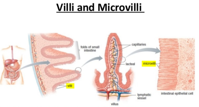
Image – Difference between Microvilli and Villi in the intestine
What are the roles or functions of microvilli?
Microvilli perform numerous functions and most of them include the following:
- They increase the cell’s surface area allowing the cell to absorb more and perform its secretory functions.
- In the intestines, microvilli work with villi to absorb all the essential nutrients by expanding the intestines’ surface area.
- The membranes of microvilli are packed with enzymes that help in breaking down complex nutrients into simpler compounds so that they can be easily absorbed by the body.
- Microvilli play an essential role in fertilization. They make fertilization easy by helping the sperm to anchor to the egg.
- Microvilli also act as an anchoring agent in white blood cells. They make white blood cell migration possible.
- Microvilli store membrane and microfilament materials.
- Microvilli also perform motility function. they can sweep unwanted materials towards the resorptive part of the cell.
- Other functions of microvilli are secretion, absorption, adhesion of cells, and mechanotransduction. (5, 6, 7, and 8)

Image 3: A comparison image between cilia and microvilli.
Picture Source: easybiologyclass.com
Difference between Cilia and Microvilli
Where they occur?
- Cilia – They are found in the columnar epithelial cells of the tubular areas of the uterine and respiratory system.
- Microvilli – They are abundant in the columnar epithelial cells of the tubules of the kidneys and small intestines.
Where they arise?
- Cilia – They arise from the basal granules
- Microvilli – They don’t arise from the basal granules.
Nature of functions
- Cilia – Primarily involved in the cellular movement.
- Microvilli – They are primarily involved in absorption.
Motility
- Cilia – They are motile.
- Microvilli – They are non-motile.
Structure
- Cilia – They consist of microtubules.
- Microvilli – They consist of microfilaments. (8, 9, and 10)
Presence of glycocalyx layer
- Cilia – They do not have a glycocalyx layer.
- Microvilli – They are surrounded by layers of a glycocalyx.
Shapes
- Cilia – They are distally tapered.
- Microvilli – They are characterized by their thin and short stature. They are cylindrical in shape with blunt ends. (3, 5)
Number
- Cilia – They are fewer in numbers when compared to microvilli.
- Microvilli – They are plenty in numbers.
Etiology
- Cilia – It is a Latin word which means eyelashes.
- Microvilli – It comes from the Greek word mikros which means small and villus, a Latin word which means hair. (3, 5, and 9)
Refer to the table below for a brief comparison between cilia and microvilli.
| Point of comparison | Cilia | Microvilli |
| Location | Found in the columnar epithelial cells of the uterine tubules and respiratory tract. | Found in the columnar epithelial cells of the kidney tubules and small intestines. |
| Where they arise | Basal granules | Do not arise from the basal granules |
| Main functions | Movement | Absorption |
| Motility | Motile | Non-motile |
| What are they made of | Microtubules (9+2) ultrastructure | Microfilaments but lack (9+2) ultrastructure |
| Presence of glycocalyx layer | Does not have glycocalyx layer | Surrounded by glycocalyx layer |
| Shapes | Distally tapered | Very thin and short/cylindrical with blunt ends |
| Number | Few in numbers | Numerous |
| Etiology | A Latin word for eyelashes | A Latin word for small hair |
Microvilli destruction
Certain diseases can cause microvilli destruction and it is linked to the host cell’s cytoskeleton rearrangement. When this happens, nutrient malabsorption takes place leading to persistent osmotic diarrhea and fever.
It is a common scenario in infections caused by E. coli such as in the case of Celiac disease and some forms of a genetic disease characterized by a defect in microvilli such as microvillus inclusion disease.
There are instances when microvilli’s destruction is all the more beneficial such as when getting rid of microvillus on white blood cells, which is essentially helpful in fighting autoimmune-related diseases. (1, 4, 7, and 10)
Quick facts about microvilli
- Microvilli are cell’s minute microscopic projections. They are present in, on, and around the cells.
- Microvilli exist on their own or with villi.
- They are a hair-like structures.
- They give the human cell 600 times more surface area.
- There are microvilli in the tongue, specifically in the taste buds responsible for sending signals to the brain telling the brain what a particular food tastes like.
- Microvilli are abundant in the intestines and their primary goal is to absorb nutrients from the digested foods. (3, 6, and 10)
References
- https://en.wikipedia.org/wiki/Microvillus
- https://link.springer.com/referenceworkentry/10.1007%2F3-540-29623-9_4120
- https://study.com/academy/lesson/microvilli-definition-function.html
- https://microbenotes.com/microvilli-structure-and-functions/
- https://mmegias.webs.uvigo.es/02-english/5-celulas/ampliaciones/7-microvellosidades.php
- https://www.sciencedirect.com/topics/engineering/microvilli
- https://www.ncbi.nlm.nih.gov/pubmed/20607764
- https://www.histology.leeds.ac.uk/tissue_types/epithelia/epi_specialisations.php
- https://www.annualreviews.org/doi/abs/10.1146/annurev-cellbio-100814-125234?journalCode=cellbio
- https://phys.org/news/2018-09-microvilli.html