MHC stands for Major Histocompatibility Complex; it is the region in the genes responsible for transplantation antigens, which are involved in shielding the body from pathogens.
MHC molecules have two major classes: MHC I and MHC II. Both have a and b chains from various sources. Let us discuss in details the MHC classes. (1, 2)
MHC class I Protein
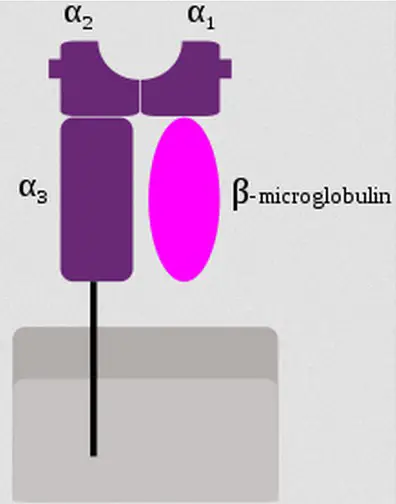
Image 1: MHC Class I protein.
Picture Source: wikimedia.org
The cells in the body, almost all of them contains MHC class I. it presents endogenous antigens from the cytoplasm. These include self-proteins as well as proteins (foreign) produced inside the cell. Once the proteins degrade, the fragments of the peptide are moved to the endoplasmic reticulum and bind to MHC I proteins.
After which, they will be transported to the cell’s surface. Once they reach the surface of the cell, the MHC I protein will display an antigen to be recognized by cytotoxic T cell lymphocyte, which is a special immune cell.
MHC I protein presents the kind of proteins that is synthesized in the cell. The killer T cells monitor such activities so as to identify and immediately get rid of over-abundant cells and foreign peptide antigens like malignant cells and viruses. (1, 2, 3, and 4)
MHC class II Protein
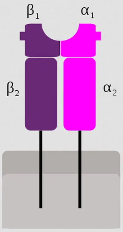
Image 2: MHC Class II Protein.
Picture Source: wikimedia.org
Unlike MHC I protein which is present in almost all cells, MHC class II protein can only be found on specialized antigen-presenting immune cells like:
- Macrophages – They engulf foreign bodies like harmful bacteria.
- Dendritic cells – They are antigen present in T cells.
- B cells – They produce antibodies.
Once the pathogenic organism is encountered, the protein from the pathogen degrades into fragments of the peptide by antigen-presenting cells and these fragments will be sequestered into endosome and bind to MHC II proteins. After which, it will be transported to the surface of the cells. Once it reaches the cell’s surface, MHC II proteins will display the antigen to be acknowledged by T cell lymphocyte.
T cell lymphocytes are helper cells and they are activated once bind to dendritic cell MHC II antigen and/or macrophage which will result in the production of lymphokines.
They will entire other cells to migrate to the infection site so as to get rid of the antigenic materials. As T cells bind to B cell MHC II antigen, they stimulate the production of clone cells (antibody-producing) in fight of antigenic materials. (3, 4, and 5)
MHC Class III
This is a special class composed of diverse complement components of genes such as Bf, C2, and C4. These are found in between class I and Class II genes and later on grouped as MHC class III.
Some genes encoded in the telomeric end of the Class III region are somewhat involved in both specific and global inflammatory responses. Molecules that belong to MHC class III also have immune-related functions. (5, 6)
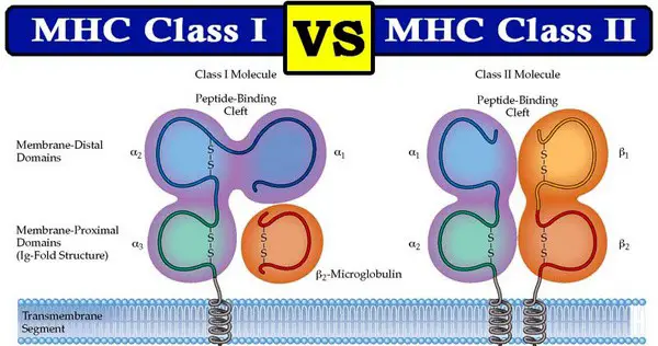
Image 3: A comparison image between MHC Class I and MHC Class II.
Picture Source: stackpathdns.com
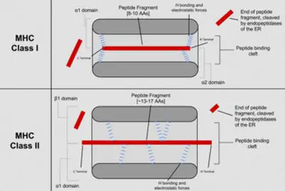
Image 4: Major histocompatibility complex class I and II difference.
Picture Source: wikimedia.org
Let us take a look at the similarities between MHC class I and MHC class II
- They are both MHC molecules encoded by clusters of MHC genes.
- They are both surface antigens expressed on the membrane of the cell.
- They are both antigens found on T cells.
- They are both involved in eliciting immune responses to fight foreign antigens.
- They are both the reasons behind graft rejection during organ and tissue transplant. (5, 6, and 7)
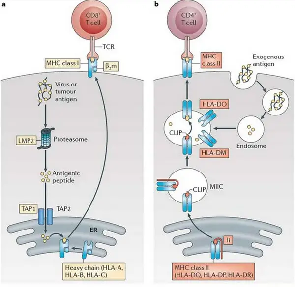
Image 5: The image shows the differences between MHC Class I and MHC Class II.
Picture Source: nature.com
What are the differences between MHC Class I and MHC Class II?
Where can they be found?
- MHC Class I – They are present on the surface of nucleated cells such as the cells in mammals.
- MHC Class II – They are present on antigen presenting cells like B cells, dendritic cells, and macrophages.
Occurrence
- MHC Class I – They are found on all nucleated cell types in the body.
- MHC Class II – They are found on antigen presenting cells like macrophages, dendritic cells, and B cells. (5, 8)
Structure
- MHC Class I – They consist of 3 alpha domains and 1 beta domain.
- MHC Class II – They consist of 2 alpha and beta domains.
Function
- MHC Class I – Their main role is to clear endogenous antigens.
- MHC Class II – Their main role is to clear exogenous antigens. (4, 6, and 9)
Encoded genes
- MHC Class I – There are three: MHC-A, MHC-B, and MHC-C.
- MHC Class II – MHC-D.
Membrane-spanning Domain
- MHC Class I – They have a single membrane-spanning alpha and beta domains.
- MHC Class II – They have two spanning alpha and beta domain membranes. (8, 9)
Antigen-presenting Domains
- MHC Class I – Both alpha 1 and 2 domains have something to do with the presentation of antigens in the molecules of MHC class I.
- MHC Class II – Both alpha 1 and beta 2 domains have something to do with the presentation of antigen in MHC Class II molecules. (2, 4)
Encoded Chromosomes
- MHC Class I – Alpha domains are found on the LHC locus of chromosome 6. On the other hand, beta chains are found on chromosome 15.
- MHC Class II – They are found on chromosome 6.
Responsive Cells
- MHC Class I – Present antigens to cytotoxic T cells.
- MHC Class II – Present antigens to helper T cells.
Nature of presenting antigen
- MHC Class I – They have endogenous antigens that came from the cytoplasm.
- MHC Class II – They have exogenous antigens that came outside of foreign organisms. (9, 10)
Responsive Co-receptor
- MHC Class I – They bind to CD8+ receptors on the cytotoxic T cells.
- MHC Class II – They bind to CD4+ receptors on the helper T cells.
Brief comparison between MHC Class I and HC Class II:
| Point of comparison | MHC Class I | MHC Class II |
| Location | Surface of nucleated cells in mammals | Antigen presenting cells like:
|
| Occurrence | Nucleated cell types in the body | Antigen-presenting cells like macrophages, dendritic cells, and B cells. |
| Structure |
|
|
| Encoded genes |
| Chromosome 6 |
| Nature of presenting antigen | Endogenous antigens that derived from the cytoplasm. | Exogenous antigens that came extracellularly from foreign organisms. |
| Antigen-presenting Domains | Alpha 1 and 2 are involved in presentation of antigens in the molecules of MHC class I. | Alpha 1 and beta 2 domains are involved in the presentation of antigen in MHC Class II molecules. |
| Responsive cells | Present antigens to cytotoxic T cells. | Present antigens to helper T cells. |
| Responsive Co-receptor | Bind to CD8+ receptors on the cytotoxic T cells | Bind to CD4+ receptors on the helper T cells |
| Function | To clear endogenous antigens | To clear exogenous antigens |
References
- http://pediaa.com/difference-between-mhc-class-1-and-2/
- https://microbeonline.com/difference-mhc-class-mhc-class-ii-proteins/
- https://www.biologyexams4u.com/2012/12/difference-between-mhc-class-i-and-mhc.html
- https://en.wikipedia.org/wiki/Major_histocompatibility_complex
- https://microbenotes.com/differences-between-mhc-class-i-and-class-ii/
- https://www.ebi.ac.uk/interpro/potm/2005_2/Page2.htm
- https://www.differencebetween.com/difference-between-mhc-i-and-vs-ii/
- https://www.immunopaedia.org.za/immunology/basics/4-mhc-antigen-presentation/
- https://www.sciencedirect.com/topics/biochemistry-genetics-and-molecular-biology/mhc-class-i
- https://courses.lumenlearning.com/boundless-microbiology/chapter/the-major-histocompatibility-complex-mhc/
