Wright’s stain is one of commonly performed staining methods that was derived from Romanowsky Stain; used to stain peripheral blood smears. Through Wright’s Staining, different blood cell types can be differentiated properly.
Among other uses and advantages are:
- Staining urine samples
- Bone marrow aspirate staining
- To check the blood smears for the presence of malarial parasites
- It is used to stain chromosomes to diagnose various diseases, infections, and syndromes such as leukemia.
The man behind Wright’s Stain is James Homer Wright. He was the man who modified the process used in Romanowsky Stain.
In 1902, Wright’s stain became an official laboratory procedure, especially in differentiating white blood cell counts, which is an integral component of evaluating blood cell morphology.

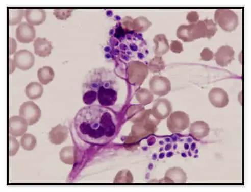
(A microscopic image of cell using Wright’s stain staining technique.)
What is the principle of Wright’s Stain?
- It is a staining procedure polychromatic in nature consist of Methylene blue and eosin mixture.
- It is methanol-based, which means it won’t need fixation step before staining.
- Fixation can do many things, such as reducing water artifacts that may take place on sunny weather or with aged stains.
- What methanol does is it ensures the cells attache to the slide.
- There are two types of dyes used in Wright’s stain; one is anion dye in the form of Eosin Y, and the other is a cationic dye in the form of methylene blue.
- Buffered water is used to dilute the solution, which triggers the process of ionization.
- Eosin is the one that stains primary components like granules of hemoglobin and eosinophilic, changing the color from orange to pink.
- The cellular components classified as acidic are stained with Methylene blue such as nucleic acid and basophilic granules in blue.
- For cell’s neutral components, they are stained by dye’s components of varying colors.
What are the reagents used?
You can buy the dye in powder form and mix it to methanol or readily available solutions.
- The staining solution consists of 1.0 gram Wright’s stain powder and 400 ml methanol (water-free).
- Phosphate buffer (pH 6.5/ 6.8, 0.15 M) – The phosphate buffer consists of 0.663 grams of potassium dihydrogen phosphate (anhydrous) 0.256 grams disodium hydrogen phosphate (anhydrous), and 100 ml distilled water.
Wright’s stain procedure
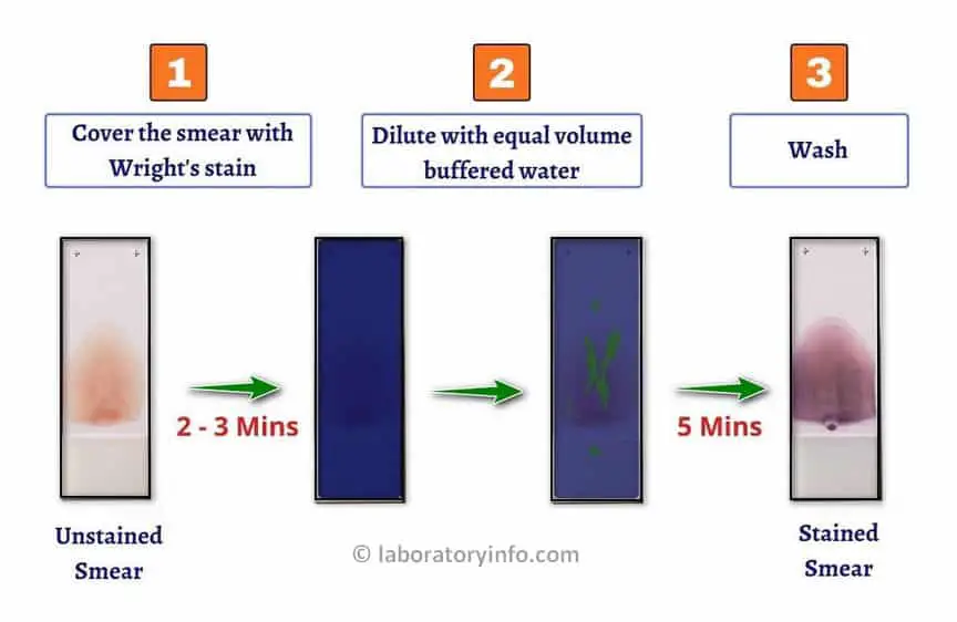
- Begin the procedure with the fixative step. Dip the slide in methanol for a few seconds and ensure that it dries up completely.
- Once the smear is air-dried completely, place it on the slide staining rack and apply 1 ml of Wright’s stain solution for about 3 minutes.
- Once all set, the next step is to add the buffer solution (phosphate) with a pH of 6.5 or exact 2 ml of distilled water.
- Allow it to stand twice for at least three minutes.
- Using water, rinse the stained smear, ensuring the pH level of the rinsing agent is 6.5. Thoroughly rinse the stained smear until the edges of the smear turn light pinkish-red.
- The underside may have some water stains; wipe it gently, and air dry in a vertical position.
- Check the slide under the microscope.

Interpreting Results
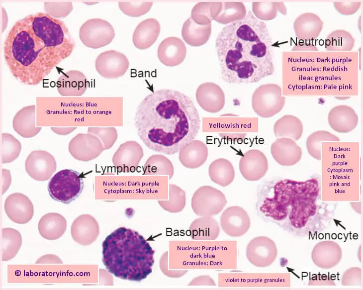
Erythrocyte – The color turns yellowish to red.
Neutrophils – The result varies depending on the specific location, such as the following:
- Granules – reddish
- Nucleus – dark purple
- Cytoplasm – pale pink
Eosinophils – For eosinophils, the following changes in colors are observed:
- Nucleus – blue
- Cytoplasm – blue
- Granules – red to orange
Basophils – For basophils, the following changes in colors can be observed:
- Nucleus – Purple to dark blue
- Granules – Dark purple
Monocytes – These changes in color are observed:
- Nucleus – dark purple
- Cytoplasm – Mosaic pink and blue
Lymphocytes – These changes in color are evident:
- Nucleus – dark purple
- Cytoplasm – Sky blue
Platelets – The granules appear violet to purple
How long does Wright’s stain last?
- Wright’s stain can last for up to 52 weeks from the manufacturing date provided stored the right way.
- It should be stored at room temperature away from direct rays of the sun.
How is Wright’s stain similar and different from Giemsa Stain?
- Wright’s stain procedure is different from other procedures because the process involves an air-dried blood film where Wright’s stain is applied and allows to dry for three minutes.
- Then, an equal amount of buffer is applied, mixed, and left for approximately five minutes.
- The slide is held in a horizontal manner and rinsed using distilled water.
- Once completely dried, the slide is examined under the microscope.

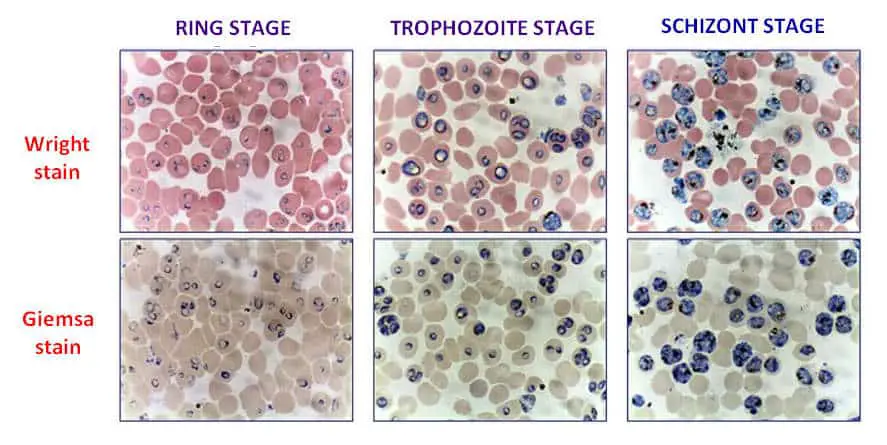
A microscopic image showing the blood films showing the various developmental stages of malaria (plasmodium falciparum) difference between Wright’s stain and Giemsa stain.
How is Wright’s stain similar to Giemsa’s stain?
- They both consist of essential components such as eosin Y, oxidized methylene blue, and azure B dyes.
- They are both used for differential staining.
- They are both essential in performing differential white blood cell counts, especially in red blood cells’ morphology study.
What are the differences between Wright’s stain and Giemsa Stain?
Although Wright’s stain and Giemsa stain are both differential staining and alike in many ways, they have differences.
For example, Giemsa stain is primarily used for bacterial cell and human cell staining, while Wright’s stain is primarily used for differentiating bone marrow aspirate, blood smears, and urine samples.
| Point of comparison | Similarities | Differences |
| Wright’s stain | – Has eosin Y, oxidized methylene blue, and azure B dyes – used for differential staining – used for white blood cell count – used for red blood cell morphology study | Primarily used for the following: – Bone marrow aspirate – Blood smears – Urine samples |
| Giemsa Stain | – Has eosin Y, oxidized methylene blue, and azure B dyes – used for differential staining – used for white blood cell count – used for red blood cell morphology study | Primarily used for bacterial cell and human cell staining. |
Conclusion
- There are different types of staining methods used in a laboratory setting. Such methods are an integral part of microscopic studies as they can significantly enhance the contrast of microscopic images.
- Wright’s stain is a sub-type of Romanowsky stain primarily involved in differential studies of white blood cell count and red blood cell morphology.
- Since it is derived from Romanowsky stain, you can expect to use the typical components like eosin Y, oxidized methylene blue, and azure B dyes.
- When it comes to differentiating components of blood smears, bone marrow aspirates, and urine samples, Wright’s Stain is the laboratory procedure of choice.
- It is essentially useful in blood cell type differentiation, especially in distinguishing various types of blood cells. Through Wright’s stain, infections can be observed and detected by observing white blood cell counts.
- Staining using Wright’s stain has been one of the most useful staining procedures.

