The Urogenital system or Urogenital system of frog is comprised of:
- Excretory System
- Reproductive System
Functionally, the excretory system is concerned with excretion of waste products and the reproductive system is concerned with producing progeny. In frogs, the reproductive organs are different in both sexes.
Excretory System of Frog
Frogs are ureotelic animals whose primary excretory product is Urea. The organs of excretion are common for both sexes. They are
- Paired Kidneys
- Ureters
- Urinary bladder
- Cloaca

Kidneys
Both kidneys are reddish brown, elongated organs of almost semilunar shape enclosed in a fibrous capsule. They lie close to the dorsal wall on the right and left side of spinal column.
Ventrally, they are covered with peritoneum. The outer border is smooth and inner border is notched in which the last one is deepest. Each notch extends outwards ventrally as a groove, each carrying a branch of portal vein.
Hence, the ventral surface is grooved and lobulated while the dorsal surface is smooth.

Image source: i.pinimg.com
Histology of Kidney:
The uriniferous tubules are the excretory units of kidneys and they originate on the posterior aspect of Malphigian corpuscle which are oval bodies comprised of loop of arterial twig with connective tissue.
There are approximately 2000 tubules in each kidneys. The Malphigian corpuscles lie close to ventral surface than dorsal surface.
The Malphigian corpuscle is composed of
- Bowmans’s capsule
- Glomerulus
The glomerulus is composed of afferent arteriole, tuft of capillaries and efferent arteriole. The blood after getting filtered in the glomerulus reach the renal vein. The capillaries arising from the efferent arteries supply the uriniferous tubules.

Image source: bszm.elte.hu
Uriniferous tubules are the functional units of kidney. These tubules are divided as 4 parts.
- First part: After its origin, the tube widens and is lined with small ciliated cubical epithelium and it runs dorsally. This is known as the neck of tubule
- Second Part: The neck is followed by a very tortuous portion on the dorsal surface and is lined with ciliated columnar epithelium. It winds ventrally.
- Third Part: This corresponds to the narrow limb of Henley’s loop and has epithelial lining similar to neck.
- Fourth Part: This corresponds to the wide limb of Henley’s loop and it ascends dorsally and open into collecting duct.

Image Source: amazonaws.com

The collecting tubule takes a horizontal course and reach the dorsal surface of kidney and open into a Bidder’s canal. The Bidder’s canal lies longitudinally on the inner surface of kidneys
Ureters
Ureters are a pair of tubes arising from the smooth outer surface of each kidneys by bifurcating branches which are in communication with the collecting ducts. On the upper two third, it lies dorsal and in the lower one third it winds to the outer aspect of kidney and continues dorsally and converge with the ureter of opposite side. They open into the dorsal aspect of cloaca through separate apertures.
In male frogs, the part of ureter behind the kidney is dilated as seminal vesicle which stores sperm. In addition to passage of urine, the ureters conduct sperms in the breeding season. Hence, they are called as urinogenital ducts or seminiferous duct. In females, the ureters are in close relation with the dilated oviducts and follow a course similar to that in male frogs.

source: brainkart.com

Image Source: brainkart.com
Urinary Bladder
The urinary bladder is a thin walled muscular structure opening into the front surface of cloaca. The opening of urinary bladder lies opposite to the ureteric apertures in the cloaca and is guarded by a mucosal fold called sphincter to prevent the back flow.
The bladder has an inner epithelial layer which rests on loose areolar tissue, middle smooth muscle layer and an outer peritoneal layer. It stores urine before it is voided out through cloaca.
Cloaca
The small sac like cloaca receives ureter, urinary bladder and rectum in the anterior part in male frogs. In females, in addition to the said apertures, a pair of oviduct opens in the posterior part. Cloaca opens to the exterior through the cloacal aperture placed between the hind limbs.
Physiology of Excretion
The blood carrying the excretory products and metabolic by products enter the kidney through via afferent arterioles in glomerulus which then drains into the portal veins of kidneys. The high renal blood pressure facilitates the glomerular filtration.
The filtrate contains plasma, urea, glucose and water and enters the uriniferous tubule through Bowman’s capsule. The filtrate goes in to the renal vein via efferent arteriole.
When the filtrate pass through the various segments of the uriniferous tubule, selective reabsorption of substances like glucose and amino acids which are recycled as energy sources and protein synthesis and abundant water and chlorides to maintain the fluid balance.
The remaining filtrate which is now concentrated with urea formed from amino acid metabolism and some salts and water pass through the ureter, stored in urinary bladder and is expelled via cloaca.
Urea is an excretory product which is less toxic than ammonia and requires less water to get excreted. Therefore, the amphibians are ureotelic in nature.
Reproductive System in Frogs
Frogs are dioceous in nature i.e., the sexes are separate. The sex organs of male and female frogs are anatomically different
Reproductive system of male frogs
The reproductive system of male frogs consists of
- Testis
- Vasa efferentia
- Urogenital or Seminiferous duct
Testis
They are located on the ventral aspect of the upper pole of kidneys and vary in shape and size based on species and season. It is spherical in its largest dimension and may be conical at times. The testis is a firm lobulated organ which presents as multiple convexities and the vessels and ducts enter and leave through the hilum of testis.
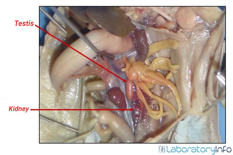
Image Source: Slideplayer.com
The testis is enclosed in a fibrous capsule that sends trabeculae within the organ and divides it as lobules. Outside the capsule, it is covered with a layer of peritoneum which extends dorsally as the double layered mesorchium and is attached to the inner aspect of the posterior body wall. Multiple finger-like projections called the Fat bodies are attached on the ventral surface of the Testis which serve as energy house for development of sperms and in the cold winters when the frogs enter hibernation.
Microscopic appearance of Testis
Each testis has an outer fibrous covering which projects within the gland and divide it as lobules. Each lobule consist of numerous seminiferous tubules which are supported by intervening connective tissues. The seminiferous tubules arise from an irregular sinus in the centre of the organ and runs to the periphery where they branch.
The seminiferous tubules are lined by two types of cell. The cells in the periphery are large, round with distinct large nucleus and occur in groups while the spermatozoa are spindle shaped cells that appear to radiate from lumen to the periphery.
Picture 9: Microscopic view of Frog Testis
The connective tissues between the lobules contain blood vessels, lymphatics and interstitial cells which secrete the androgen, Testosterone. It is a vital hormone which is responsible for sexual characteristics of male frogs like altering the contractile properties of the vocal muscles during the breeding season.
Vasa Efferentia
The vasa efferentia are multiple tubules which arise from the hilum of testis. They run inwards in the mesorchium and run dorsally on reaching the kidney and lies between kidney and testes. Then its curves ventrally and lies on the inner surface and kidney and open into the Bidders canal.
Most of the Vasa efferentia open into collecting duct via Bidders’ canal while some of them end blindly in the mesorchium. The mature sperms enter the lumen of the vasa efferentia which then enters the ureter via Bidders canal. From ureter, it is stored in the Seminal vesical which is a sac like dilation on the upper part of ureter.
Urogenital/ Seminiferous Duct
In male frogs, the Ureter is a common conduit for both urine and mature sperms and both ureters open on the dorsal aspect of cloaca through separate apertures guarded by sphincters.
Reproductive System of Female frogs
The components of reproductive system in female frogs are
- Paired ovaries
- The oviducts
Ovaries
The pair of ovaries are sac like structures which are divided to multiple lobules by thin walled septa. Similar to mesorchium of Testis in male frogs, each ovary is enclosed by a double layered peritoneal fold called the mesovarium which suspends it from the dorsal wall.
They connected to oviduct and are not in communication with the Bidders canal. Hence, female frogs have a distinct excretory and reproductive system unlike male frogs. Ovaries enlarge in size during the mating season.

Source: Springnature.com
Histology of Ovary
Both ovaries are enclosed in a layer of peritoneum lined with ciliated epithelium and a layer of connective tissue that extends inwards as the septa dividing the ovary. The egg cells or ova are attached on the inner aspect these septa. Patches of germinal epithelium lies between the peritoneal and the fibrous layer and are numerous in the periphery of the organ. The connective tissue also contain blood vessels, lymphatics and nerves.
Fig 11: Microscopic view of immature ovary of frog
The cells of the germinal epithelium forms cluster of immature egg cells called oogonia. This collection of oogonia is called ovarian follicle. Each ovarian follicle has a cell in the middle of the cluster which enlarges into the mature ovum while the remaining cells contribute to the formation of the follicular epithelium and are replaced by the vitelline membrane of the mature ovum. The cytoplasm of mature ovum contains abundant yolk granules in the cytoplasm.
The eggs are round shaped with and animal pole and a vegetal pole. The upper portion of the eggs have a black pigments and is referred as the animal pole. It contains the nucleus which becomes the future embryo. The lower half of the eggs is called the vegetal pole and is filled with yolk which nourishes the growing embryo. It is surrounded by inner vitelline membrane and outer albumin coat.
Fig 12: Unfertilized egg of frog
Oviducts
In immature frogs, the oviducts are short tubes. In mature frogs, during the breeding season, they become highly convoluted tubes and covers the entire kidneys. The openings of the oviduct lies in the pleuroperitoneal cavity close to the undersurface of lungs.
The oviduct runs down towards cloaca and form a dilatation called ovisac. The ovisac opens into the cloaca through a narrow opening on a papillae placed in the cloaca. Both oviducts open through two separate, closely placed apertures guarded by mucosal fold on which both ureters open.
The oviduct are lined by inner ciliated epithelium which direct the mature ovum into in to the ovisac where it is stored until fertilization. The middle layer is a thick glandular layer which contain Colloid granules which are capable of abundant water absorption and secretion of mucoid material to surround the egg before it reaches the cloaca. The outer layer is made of thin peritoneal membrane.
Reproduction in frog
In most of the frog species, fertilisation of egg occurs externally. During breeding season, the male frog makes the mating call and the female frog identifies it species and find the male partner.
They take a common position called Amplexus in which the male frog lies on top of the female in such a way that the ovum released by the female through the cloaca is fertilized with the sperm released by the male. Fertilization takes place externally in a moist or wet environment .

Image source: wikipedia cc

Image source: stackpathdns.com
Frequently Asked Questions
Q1. How many eggs does a female frog lay?
Q2. Why is fertilization external in frogs?
Thus a female frog cannot carry the embryos within its body. Hence the type of fertilization in frog is external.
Q3. Why do frogs lay a large number of eggs?
To ensure the survival of the progeny and for the preservation of the species, laying abundant eggs is an evolutionary advantage of the frogs.
References
- https://en.wikipedia.org/wiki/Frog
- Tesler, P. (1999). “The amazing adaptable frog”. Exploratorium:: The museum of science, art and human perception.
- The Anatomy of Frog – Alexander Ecker
- A laboratory guide to Frog’s Anatomy – Eli C.Minkoff
- The Biology of Frog – Samuel J Holmes