DNA is one of the most important structures in the body. It controls both your physical traits such as the color of the hair, eyes, the number of fingers, and the shape of the nose, to name a few.
DNA is arranged into chromosomes. Humans have a total of 23 pairs of chromosomes or 46 individual chromosomes. 23 comes from the mother and the other 23 comes from the father.
Chromosomes have thousands of genes that control many traits. If there are abnormalities in the structure and number of chromosomes, physical, developmental, and health problems could occur.
To check for chromosomal abnormalities, a special procedure called karyotyping is done. Such type of procedure can capture the chromosomes of a person and check for any damage in size, shape, and number. (1, 2, and 3)
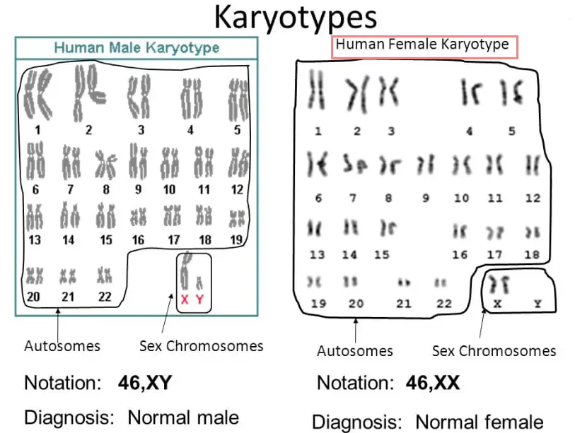
Image 1: Image of chromosomes that are arranged in chronological orders (normal : male and female ).
Picture Source: chromosome18.org
What is karyotyping?
A karyotype is a representation of the person’s chromosomes. To get a picture of chromosomes, the chromosomes are isolated, stained, and checked under the microscope.
Through the microscopic examination, a picture of the chromosomes is taken, cut up, and rearranged according to their size from largest to smallest.
What is the purpose of karyotyping?
The primary purpose of karyotyping is to look for abnormal numbers or structures of chromosomes.
Various characteristics of karyotypes are observed which include the differences in the absolute sizes of chromosomes, differences in the centromere’s position, differences in the relative size of chromosomes, differences in the basic number of chromosomes, number and position of satellites, and the differences in the degree and distribution of heterochromatic regions. (2, 3, and 4)
Chromosomal abnormalities that are detected using karyotyping are categorized into the following:
- Trisomies – There are three copies of one of the chromosomes. In a normal circumstance, there should only be two copies of a chromosome.
- Monosomies – Instead of two copies of a chromosome, there is only one copy.
- Chromosome deletions – A part of the chromosome is missing.
- Chromosomal translocations – A part of the chromosome attaches to another chromosome and vice versa. (3, 4, and 5)


Karyotyping of patient with turners syndrome
How is the karyotyping test done?
Karyotypes are done using a standardized staining procedure to reveal the structural features and characteristics of every chromosome. Human karyotypes are analyzed by clinical cytogeneticists. They are the person who checks for any genetic changes or anomalies.
Karyotyping is now used as a diagnostic procedure to check for birth defects, genetic disorders, and some types of cancer.
Karyotyping – Video Procedure (Animation)
Below video : Karyotyping procedure in animation (sorry for the bad audio quality)
Below Video : Making chromosomes spread for karyotyping
Karyotyping during pregnancy
If you are pregnant, your doctor will perform different types of procedures as a part of prenatal screening. The purpose is to check for genetic and chromosomal abnormalities. It is usually ordered during the first trimester and second trimester.
The expected result should be normal and no further test is needed. However, if there are abnormalities detected, your doctor might order another set of test to further confirm the diagnosis. The doctor needs a small sample of the baby’s cell to examine the chromosomes.
During the karyotyping procedure, the baby’s chromosomes are examined to check for any abnormalities. Under normal circumstances, the human chromosomes have a total of 23 pairs leading to a total number of 46 chromosomes. 23 of the chromosomes come from the mother and the other 23 come from the father.
In an abnormal circumstance, the baby could have a missing chromosome or an extra chromosome. Any abnormalities in the structure and number of chromosomes can lead to abnormalities in the baby. (4, 5, 6, and 7)
Abnormalities that can be detected using karyotyping procedure include the following:
#1 – Down Syndrome/Trisomy 21
A trisomy 21 or popularly known as Down syndrome is a chromosomal condition wherein the baby has an extra copy of chromosome 21. So, instead of two copies, the baby has three copies of chromosome 21 leading to the development of Down syndrome symptoms which include the following:
- Flat nose
- Small ears
- Mild to moderate issues with thinking, understanding, and reasoning.
- Delayed social skills
- Developmental milestones like walking and talking may take some time to achieve.
- Trouble hearing, seeing, and the potential to develop heart-related problems. (5, 7, and 8)

Image 2: A patient with Trisomy 21 or Down syndrome.
Picture Source: americanpregnancy.org
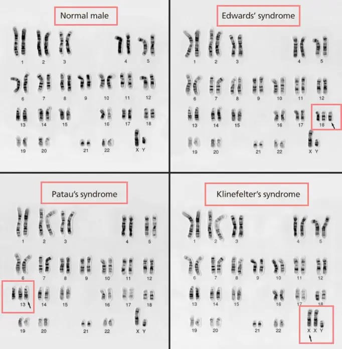
#2 – Edward’s syndrome/Trisomy 18
The condition is called trisomy 18 because the baby has extra 18th chromosomes. It is caused by an error in cell division, specifically, a meiotic disjunction. This chromosomal abnormality is life-threatening even before birth. It is extremely rare though, as one out of every 2500 pregnancies in the United States and 1 in 6,000 live births.
- Slow growth before birth
- Low birth weight
- Heart defects
- Small, abnormally shaped head
- Small mouth and jaw
- Clenched fist with overlapping fingers (3, 5, 8, and 9)
#3 – Patau Syndrome/Trisomy 13
This chromosomal abnormality is caused by an extra 13th chromosomes. Babies born with trisomy 13 won’t live more than a year. they could also have severe problems like heart-related problems and mental impairment.
- Severe intellectual disability
- Brain/spinal cord abnormalities
- Poorly developed eyes
- Cleft lip and palate
- Extra fingers or toes
- Weak muscle tone (2, 8, and 10)

Image 4: A person with trisomy 13 or Patau Syndrome.
Picture Source:wikimedia.org
#4 – Klinefelter Syndrome
It affects the male population. Male gender has XY chromosomes but what happens in Klinefelter syndrome is that the baby has an extra X chromosome. So, instead of XY, the baby has XXY. Babies with Klinefelter syndrome may undergo puberty at a slow pace. He may not have the capability to have his own children.
- Small testes that produced a small amount of testosterone
- Breast enlargement
- Decrease bone density
- Decrease muscle mass
- Reduced amount of body and facial hair
- Abnormal fusion of some bones in the forearm
- Flat feet
- Curved pinky fingers (3, 6, and 8)

Image 5: An adult who suffers from Klinefelter syndrome.
Picture Source: i.ytimg.com
#5 – Turner Syndrome
It is a chromosomal abnormality affecting females. Its either there is a missing or a damaged X chromosome. Clinical features of Turner syndrome include the following:
- Short stature
- Early loss of ovarian function
- The majority of girls with Turner syndrome do not undergo puberty unless they undergo hormone therapy.
- Webbed neck (extra folds of skin on the neck)
- Low hairline at the back of the neck
- Swelling of the hands and feet
- Skeletal-related problems
- Kidney-related problems
- Heart defects
- Possible developmental delays, behavioral problems, and non-verbal learning disabilities. (1, 4, 7, and 10)

Image 6: Physical traits of a person living with Turner syndrome.
Picture Source: medicalhomeportal.org


Image 7: The image above shows how a chorionic villus sampling procedure is done.
Picture Source: extranet.who.int
Image 8: Amniocentesis is another way of checking for chromosomal abnormalities.
Picture Source:scientificanimations.com
What do the results mean?
Karyotyping can check for any chromosomal abnormalities, specifically, extra chromosomes, missing chromosomes, extra or missing part of the chromosomes, and parts that have broken off of one chromosome and reattached to another chromosome.
The laboratory technician can check the size, shape, and number of chromosomes. The results are definite. It’s either your baby has chromosomal defects or not.
The normal result would show a total number of 46 chromosomes. Out of 46 chromosomes, two are sex chromosomes and the rest are autosomes. Abnormalities in the result show chromosomal/genetic abnormalities. However, the test should be repeated to confirm the result as there are instances when the abnormality will occur in the lab sample and not really in the body. (3, 6, 9, and 10)
Drawing samples for karyotyping procedure
#1 – Drawing blood samples from the vein
- The flow of blood is stopped by wrapping an elastic band around the upper arm. It enlarges the vein below the band making it easier to extract a blood sample.
- The insertion site is cleaned using alcohol.
- The needle is inserted into the vein and a tube is attached to the needle and fill with blood.
- The band is removed once adequate blood sample has been collected.
- The needle is removed and a cotton is pressed against the site to stop the bleeding.
#2 – Drawing sample from the fetus
Collecting samples from the fetus can be done in two ways – amniocentesis and chorionic villus sampling.
CVS/Chorionic Villus Sampling
A sample of the baby’s cell is removed from the chorionic villi using a long needle. The chorionic villi are tissues found within the placenta. The sample cell is sent to the laboratory for examination.
Depending on the result, it could show if your baby has chromosomal defects such as trisomy 21 or down syndrome, trisomy 13, trisomy 18, and other types of chromosomal defects. The CVS or chorionic villus sampling is usually done between 10 and 13 weeks of pregnancy.
However, there are some risks involved in doing the procedure such as the chance of miscarriage. The probability is 1 out of every 100 women and babies.
Amniocentesis
In this type of procedure, the doctor takes a small amount of amniotic fluid using a long needle. The needle is stick through the abdomen and the sample is sent to the lab for further examination.
Karyotyping using amniocentesis can detect not only chromosomal abnormalities but also neural tube defects; medical conditions that affect the brain and the spine. Amniocentesis is done between 15 and 20 weeks of pregnancy.
Miscarriage may happen but the possibility is 1 out of every 200 women. (1, 2, 8, and 10)
#3 – Drawing blood sample from the bone marrow
A bone marrow aspiration can also be used for karyotyping procedure.
How much is karyotyping procedure?
The cost of karyotyping varies depending on the site where the sample is taken. It usually ranges between $200 and $350.
References
- https://www.nature.com/scitable/topicpage/karyotyping-for-chromosomal-abnormalities-298/
- https://www.healthline.com/health/karyotyping
- https://www.webmd.com/baby/what-is-a-karyotype-test#1
- https://study.com/academy/lesson/karyotyping-tests-definition-procedure-examples.html
- https://www.labtestsonline.org.au/learning/test-index/chromosome-analysis-karyotyping
- https://en.wikipedia.org/wiki/Karyotype
- https://www.verywellhealth.com/how-to-how-is-a-karyotype-test-done-1120402
- https://medlineplus.gov/ency/article/003935.htm
- https://www.jove.com/science-education/10787/karyotyping
- https://www.uofmhealth.org/health-library/hw6392