Introduction
Animal cells are eukaryotic cells, mostly multicellular containing cytoplasm and membrane-bounded organelles enclosed within the plasma membrane. The animal kingdom contains the largest number of species on the entire earth.
Animals are heterotrophic organisms that contain various organelles and systems to break down the food obtained from plants or animal resources.
These organisms are motile. However, a few of them, like a hydra, are sessile. They have communication systems to communicate with other animals. Certain immune responses protect animals from invading microbes.
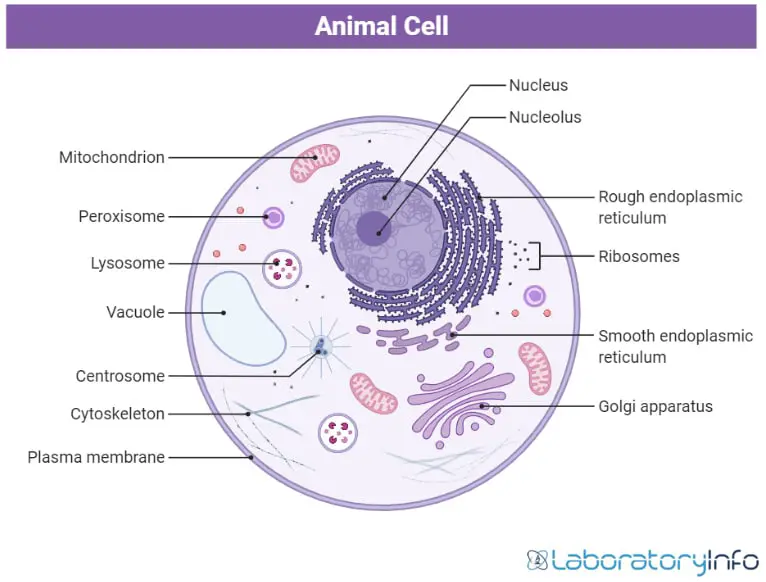
Image created with the help of biorender
Difference between Prokaryotic Cell and Eukaryotic Cell
Prokaryotic cells are unicellular that do not contain membrane-bounded organelles. Their nucleic material is dispersed in the cytoplasm. On the other hand, eukaryotic cells are multicellular, containing the membrane-bounded organelles. Their nucleic material is enclosed in the double nucleic membrane.
There are two types of cells in the bodies of organisms.
Structural cells
These cells build up the skeleton of cells to provide support to the organs—for example, epithelial cells.
Functional cells
Functional cells perform essential functions for the survival of an organism, for example, nerve cells. All living organisms are made up of various types of cells. However, there are a few differences between the structure and organelles of the cells found in different organisms,
Difference between Animal Cell and Plant cell
Both plants and animals have eukaryotic cells. However, there are a few differences between both of them. Plant cells have an additional membrane called a cell wall outside their plasma membrane. While in animal cells, the cell wall is absent. It makes plant cells more rigid than plant cells.
Chloroplast, an essential organelle containing photosynthetic pigments in plants, is absent in animal cells. The ability to move and complex organ systems make animals more complex than plants.
Image created with the help of biorender
Animal Cells
Animal cells are of various shapes and sizes. Their sizes range from micrometers to a few inches. The smallest cell in the animal is a neuron that is just 100 micrometers in diameter. Likewise, the largest cell is the egg of an ostrich that can be up to 150 millimeters in diameter and weigh about 3 pounds.
Due to the absence of cell walls, animal cells are less rigid and can take up various shapes. They can be oval, circular, spindle-shaped, and so on. Most animals’ cells are so small that they cannot be seen with the naked eye. They are usually stained to observe under a light microscope.
All the organelles are enclosed in the membrane and accomplish various functions like digestion, cellular energy production, protein synthesis, etc., that are vital for survival. Some important structures in the animals are:
Plasma Membrane
The plasma membrane is a semipermeable membrane composed of phospholipids and proteins that control the entry of different kinds of materials in the cell by allowing selective material to pass through it. The proteins are embedded in the lipid bilayer in a mosaic manner. It is present in all types of cells, whereas the composition may vary for each cell.
Small molecules like O2, CO2, and water pass easily through the phospholipids. Some other nutrients enter the cell through the protein channels in the plasma membrane. Plasma membrane encloses all the materials in the cell and serves as sites for various processes to occur.

Nucleus
The nucleus is also a double membranous organelle containing the genetic material enclosed in the nuclear envelope. It is present in the center of animal cells. Like red blood cells, some animal cells lose their nucleus upon differentiation.
It contains the genetic material called chromatin which condenses to form a chromosome. The number of chromosomes is fixed in all the species. Genes on the chromosomes give the characteristic features to a specie. The nucleus also contains nucleolus, that is, ribosome factories. Different materials enter and leave the nucleus through the nuclear pores.

Image created with Biorender
Cytoplasm
The cytoplasm is a crystal-colloidal material that contains a wide range of organelles. It is a site of occurrence for various metabolic processes. For example, glycolysis occurs in the cytoplasm of the cell. Major organelles present in the cytoplasm are the endoplasmic reticulum, ribosomes, Golgi apparatus, lysosomes, etc.

Image created with Biorender
Mitochondria
Mitochondria is present in all types of cells except for the blood cells. These are called the cell’s powerhouse as they are the major sites of ATP (cellular energy) synthesis in the cell. The largest number of mitochondria is present in the muscle cells. These are double membranous, with the outer being smooth and the inner layer containing gel-like matrix forms various folding called cristae.
This organelle is self-replicating as it contains its DNA. It is interesting to know that human inherits their mitochondria from the mother as it comes from the egg cell after fertilization. Kreb cycle and electron transport chain that are the major steps in the energy generation in the cellular energy production occur in the mitochondria of the cells.

Image created with Biorender
Ribosomes
Ribosomes are the protein factories containing 60% RNA and 40% proteins. These are smaller and can be free-floating in the cytoplasm or attached to the endoplasmic reticulum. The smaller subunit (the 40S) and larger subunit (50S) together form 80S units in the eukaryotes.
Ribosomes are essential in the central dogma in the process of translation. In translation, proteins are formed by binding amino acids according to the sequence encoded in the mRNA. After the completion, a smaller and larger subunit of the ribosome separates, and protein is released.

Image created with Biorender
Endoplasmic Reticulum
The endoplasmic reticulum is the organelles with folded membranes present near the nucleus in the cytoplasm. They are of two types.
Rough endoplasmic reticulum
The rough endoplasmic reticulum contains ribosomes and is the site of protein synthesis.
Smooth endoplasmic reticulum
The smooth endoplasmic reticulum does not have ribosomes and performs functions like the formation of membranes, steroid production, and detoxification of the cells.

Image created with Biorender
Golgi Apparatus
Golgi apparatus is present near the endoplasmic reticulum and consists of flattened sacs known as cisternae. The material from the endoplasmic reticulum enters the Golgi apparatus, modified and packed before passing them to the sites where they are required. Modification can be of various types, like breaking down complex molecules like oligosaccharides, adding or removing different components, etc.
The two faces of Golgi bodies are the cis face and trans face. Materials enter the Golgi apparatus to the cis face and leave it from the trans face after modification.

Lysosome
Lysosomes are single membranous organelles that contain different types of digestive enzymes. One of the major roles of the lysosome in the cell is autophagy to remove old parts of the cells. These are acidic due to the hydrolytic enzymes present in them.
The lysosome function is to break down different macromolecules into micro molecules and renew the cell parts.

Image created with Biorender
Cytoskeleton
The cytoskeleton is a protein structure responsible for giving proper shape to the cell and helping in mitosis. These are of three types.
Microfilaments
Microfilaments are composed of actin proteins and give shape to the cells and help in the movement.
Microtubules
These are composed of tubulin protein and are essential in forming spindle fibers for cell division.
Intermediate filaments
These are stable filaments and give elasticity and proper structure to the cells.

Image created with Biorender
Centrioles
Centrioles are composed of 9 microtubule triplets. During the cell division, centriole replicates, and two of them move to the opposite poles and start forming spindle fibers. The function of the spindle fibers is to push two daughter chromosomes apart to form two daughter cells from a single parent cell.

Image created with Biorender
Peroxisome
As its name indicates, peroxisomes are small organelles that produce hydrogen peroxide in the cells. Hydrogen peroxide neutralizes various toxic materials present in the cells. These are also involved in lipid metabolism.

Image created with Biorender
Vacuoles
The vacuoles are regulatory organelles enclosed in a single membrane called a tonoplast. There are several small vacuoles present in the animal cells, unlike plant cells, in which only one large vacuole is present. They store water, nutrients, and waste material to be removed from the cell or move inward.

Image created with Biorender
Frequently Asked Questions
Q1. What are the animal cells?
Q2. What are the few processes that occur in mitochondria?
Q3. Where does the electron transport chain occur?
Q4. Why is the rough endoplasmic reticulum called so?
Q5. In which organelle modification of materials occur?
References
- Microbiology: An Introduction 12th Edition by Gerard J. Tortora
- Molecular Biology by Robert F.Weaver fifth edition
- Bruce Alberts: Molecular Biology of the Cell Sixth edition
- https://microbenotes.com/animal-cell-definition-structure-parts-functions-and-diagram/
- https://micro.magnet.fsu.edu/cells/animalcell.html#:~:text=Animal%20cells%20are%20typical%20of,not%20have%20a%20cell%20wall.
- https://byjus.com/biology/animal-cell/