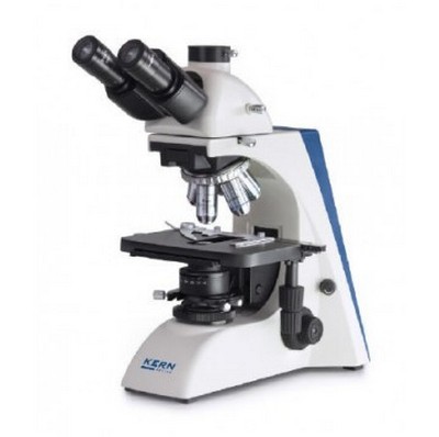
A microscope is a laboratory instrument used to see things that are not visible to the human eyes. There are different types of microscopes and each type has its benefits and advantages. These are the following:

Image 1: A simple microscope is the first microscope ever created.
Picture Source: stackpathdns.com

Image 2: An image of a compound microscope.
Picture Source: imimg.com
#1 – Types of microscopes according to lenses
- Simple microscope – it was the very first microscope created by Antony Van Leeuwenhoek in the 17th century. It was a magnifying glass, so simple yet powerful and useful. It consists of a single lens or lenses group in one unit.
- Compound microscope – it is called a compound microscope because it has two lenses resulting in better magnification. It is a bright field microscope that provides up to 1,000 times magnification. (1, 2, 3, and 4)
Light microscope/Optical Microscope
This type of microscope uses light to produce the viewed image. Light microscopes have various sub-types which include the following:

Image 3: A basic single magnification microscope.
Picture Source: olympus-lifescience.com
- Basic single magnification microscope – It is similar to that of the magnifying glass with a magnification power of 20x. It uses ambient light to illuminate the specimen being examined. This microscope is used to viewing objects like bugs, fabric weaves, and sand grains.

Image 4: A basic introductory biological compound microscope/transmitted light microscope.
Picture Source: balance-express.com
- Basic introductory biological compound microscope/transmitted light microscope – it is usually used in elementary and middle schools. It comes with three objective powers: 40x, 100x, and 400x. Beneath the stage is where light is located and where the slide is placed.

Image 5: A full-size biological compound microscope.
Picture Source: walmartimages.com
- Full-size biological compound microscope – It comes with three to four magnifications: 40x, 100x, 400x, and 1000x. The light is under the stage to transmit illumination. It comes with a built-in mechanical stage enabling the students to easily maneuver the slides on the stage.

Image 6: An example of a shop microscope.
Picture Source: walmartimages.com
- Shop microscope – It has a small flashlight used to illuminate specimens. It is placed directly on the specimen with the light on. This type of microscope is commonly used in the publishing industry to examine printed materials. It is also useful in the textile industry, specifically in checking the color and weaves of the fabric.

Image 7: An example of a binocular research microscope.
Picture Source: imimg.com
- Research microscope – it comes with fluorescence and primarily used by cell biologists. In fact, it is the microscope of choice when localizing proteins in a sample.

Image 8: The image depicts an inverted microscope.
Picture Source: imimg.com
- Inverted microscope – It is used in biology setting and comes with a light on top and objective lenses beneath the stage. It is specially created for transmitted light observations. Its magnification powers are 40x, 100x, 200x, and 400x.

Image 9: The image is one of the classic models of a metallurgical microscope.
Picture Source: microscopeworld.com
- Metallurgical microscope – It is a high power microscope with reflected light illumination, which enables you to view materials that typically won’t allow light to pass through such as plastic and metal. Its magnification ranges from 40x to 1000x.

Image 10: A polarizing microscope.
Picture Source: microscope.com
- Polarizing microscope –It uses polarizer and analyzer to visualize materials under polarized light. Specimens can be viewed spectacularly under polarized light. A polarizing microscope is commonly used in industries like geology and pharmaceuticals.
- Brightfield microscope – It uses transmitted light to check for a target at maximum magnification.

Image 12: A Phase contrast microscope uses a light interference.
Picture Source: shopify.com
- Phase contrast microscope – It examines very tiny surface irregularities using light interference. It has the ability to check living cells without having the need to stain them.

Image 13: A differential interference contrast microscope is a huge type of microscope that uses polarized light.
Picture Source: olympus-lifescience.com
- Differential interference contrast microscope – it is somewhat similar to the face contrast in terms of observing even the smallest surface irregularities. It uses polarized light which can limit the different observable specimen containers.

Image 14: A fluorescence microscope uses a special light source.
Picture Source: spachoptics.com
- Fluorescence microscope – it observes fluorescence emitted by the sample with the use of a special light source such as a mercury lamp.

Image 15: This is what a total internal reflection fluorescence microscope looks like.
Picture Source: biocrf.ust.hk
- Total internal reflection fluorescence microscope – It is somewhat the same as the fluorescence microscope instead that it uses the evanescent wave to illuminate the surface close to the specimen.

Image 16: A laser microscope is also known as a laser scanning confocal microscope.
Picture Source: itn-snal.net
- Laser microscope – It uses laser beams to clearly observe thick samples with different focal distance.

Image 17: A structured illumination microscope.
Picture Source: meyerinst.com
- Structured illumination microscope – A microscope with a high resolution and advanced technology, which has the ability to overcome limited resolution a light/optical microscope usually have caused by diffraction of light.

Image 18: A multiphoton excitation microscope from Olympus.
Picture Source: biocompare.com
- Multiphoton excitation microscope – it uses multiple excitation lasers which help reduce the damage to the cells and enabling high-resolution observation of deep areas of the specimen. In a clinical setting, the multiphoton excitation microscope is used to visualize nerve cells and the flow of blood in the brain. (2, 3, 4, 5, 6, and 7)
Refer to the table below for the summary of different types of light microscope/optical microscope.
| Types of light microscope/optical microscope | Definition |
| Basic single magnification microscope |
|
| Basic introductory biological compound microscope/transmitted light microscope |
|
| Full size biological compound microscope |
|
| Shop microscope |
|
| Research microscope |
|
| Inverted microscope |
|
| Metallurgical microscope |
|
| Polarizing microscope |
|
| Brightfield microscope |
|
| Phase contrast microscope |
|
| Differential interference contrast microscope |
|
| Fluorescence microscope |
|
| Total internal reflection fluorescence microscope |
|
| Laser microscope |
|
| Structured illumination microscope |
|
| Multiphoton excitation microscope |
|
#3 – Electron microscope

Image 19: A scanning electron microscope (SEM)
Picture Source: cloudfront.net

Image 20:A transmission electron microscope.
Picture Source: hitachi-hightech.com
It uses an electron beam as the source of light. To be able to generate the image being viewed, it needs to use computer software. Its resolution is way better when compared with the light microscope, which is perfect for seeing the inside structure of the cell. There are different types of an electron microscope and these are:
- Scanning electron microscope (SEM) – It bounces the electrons off the object thereby creating a 3D image. Its maximum magnification is 100,000x.
- Transmission electron microscope – It produces a 2D image with a maximum magnification of 500,000x enabling to visualize the inside structure of the cell. (4, 5, 7, 8, and 9)
| Types of electron microscope | Definition |
| Scanning Electron Microscope (SEM) |
|
| Transmission Electron Microscope |
|
#4 – Stereo Microscope/Dissecting microscope
It is an optical type of microscope enabling the viewer to see the sample in 3-dimensions. Its magnification power ranges from 10x to 80x. It is primarily used to inspect large specimens like fossils, rocks, coins, hair follicles, stamps, parts of flowers, and the likes.

Image 19: A gemological microscope is perfect for examining gems and minerals.
Picture Source: shopify.com
- Gemological microscope – A stereo microscope that has a special illumination perfect for examining gems and minerals.

Image 20: A fixed stereo microscope.
Picture Source: microscopegenius.com
- Stereo fixed microscope – It has fixed magnification using two objective lenses. The magnification has a fixed degree and is limited by the lens capability. To increase the magnification, you need to change the eyepiece.

Image 21: A stereo turret microscope is a more affordable option for a stereo microscope.
Picture Source: microscopeworld.com
- Stereo Turret Microscope – It comes with various mountings and one of which is the turret style. This type of mounting indicates an additional objective lens which you can rotate to your viewing position. The viewer can easily change the magnification by rotating the mounting of a turret. It is preferred by many because it is more affordable than other types of stereo microscope.

Image 22: This is how a stereo zoom microscope looks like.
Picture Source: microscopeworld.com
- Stereo Zoom Microscope – The most popular type of stereo microscope. You can easily zoom in and out to achieve the desired magnification. You can have a clear magnification by changing the eyepiece. (6, 9, 10, 11, and 12)
| Types of stereo microscope/dissecting microscope | Definition |
| Gemological microscope | It is used to examine gems and minerals. |
| Stereo fixed microscope | It has fixed magnification with two objective lenses. |
| Stereo Turret Microscope | It is preferred by many because it is more affordable than other types of stereo microscope. |
| Stereo Zoom Microscope | It is the most popular type of stereo microscope. |
| Changing the eyepiece gives you a clear magnification. |
#5 – Scanning Probe Microscope (SPM)

Image 23: A typical look of a scanning probe microscope.
Picture Source: imimg.com
A physical probe is used close to the sample so as to generate a micrograph. Examples of scanning probe microscopes include:
- Scanning tunneling microscope
- Atomic force microscope
- Magnetic force microscope
- Electric force microscope
- Near-field scanning optical microscope (12)
#6 – Point Projection Microscopes

Image 24: A point projection microscope.
Picture Source: imimg.com
A type of microscope in which ions are excited from the needle-shaped specimen and hit a detector. Examples of point projection microscopes are:
- Field emission microscope
- Atom probe microscope
- Field ion microscope (13)
#7 – Acoustic Microscopes

Image 25: An acoustic microscope
Picture Source: kibero.com
A microscope that uses sound waves to create an enlarged image of a small object. It is used to detect defects in sub-surfaces of materials.
Type of microscope according to structure
- Upright microscope – It examines the subject from above. A specimen is put on a slide and examined microscopically.
- Inverted microscope – It examines the subject from below. It is the instrument of choice for observation of samples like cells soaked with culture in a Petri dish. (12, 14, and 15)
| Microscopes according to structure | Differences |
| Upright microscope | It examines the subject from above. |
| Inverted microscope | It examines the subject from below. |
#9 – Other types

Image 26: The image above is a digital cordless microscope.
Picture Source: bhphotovideo.com
- Digital microscope – It uses optical lenses and CMOS/CCD sensors. What’s good about digital microscope is that it has a maximum magnification power of 1000x. It has the ability to record a high-quality image of the specimen. A CCD camera is attached to the microscope and connected to the LCD monitor or computer.

Image 27: A dark field microscope enables the light object to be seen on a dark background.
Picture Source: slidesharecdn.com
- Dark-field Microscope – It is used to examine live spirochetes. It comes with a special condenser lens that facilitates scattering of light. The light is reflected off the specimen at an angle. Hence, a light object can be seen on a dark background. (10, 11, 13, and 14)
|
Other types of microscopes | Differences |
| Digital microscope | Uses optical lenses and CMOS/CCD sensors. |
| Has a maximum magnification power of 1000x. | |
| Can record a high-quality image of the specimen. | |
| Dark field microscope | Used to examine live spirochetes. |
| Has a condenser lens to facilitate scattering of light. | |
| It enables light objects to be seen on a dark background. |
References
- https://www.microscopemaster.com/different-types-of-microscopes.html
- https://sciencing.com/different-kinds-microscopes-uses-5024481.html
- https://www.cas.miamioh.edu/mbiws/microscopes/types.html
- https://www.keyence.com/ss/products/microscope/bz-x/study/principle/type.jsp
- https://www.cliffsnotes.com/study-guides/biology/microbiology/microscopy/types-of-microscopes
- https://www.microscope-detective.com/types-of-microscopes.html
- https://en.wikipedia.org/wiki/Microscope
- https://eco-globe.com/types-of-microscope/
- https://owlcation.com/stem/light-and-electron-microscopy
- https://sciencestruck.com/types-of-microscopes-their-uses
- https://microscope-microscope.org/microscope-info/microscope-types/
- https://www.britannica.com/technology/microscope
- http://www.biologydiscussion.com/biology/5-important-types-of-microscopes-used-in-biology-with-diagram/2635
- https://study.com/academy/lesson/types-of-microscopes-election-light-fluorescence.html
- http://www.edinformatics.com/inventions_inventors/microscope.htm

